
eduo
visual
Cardiovascular
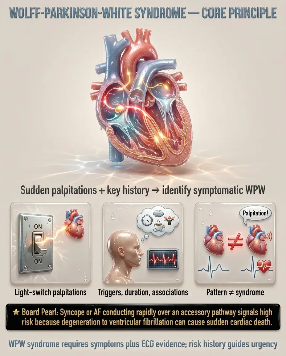
Wolff-Parkinson-White syndrome
Wolff-Parkinson-White (WPW) syndrome = presence of an accessory pathway (bundle of Kent) that creates a direct electrical connection between the atria and ventricles, bypassing the AV node → ventricular pre-excitation.
— Resting ECG shows short PR interval (<120 ms), delta wave, widened QRS
— Recurrent episodes of paroxysmal SVT in a young patient
— Irregular wide-complex tachycardia (pre-excited AF) — a life-threatening presentation
Board pearl: WPW is the most common cause of pre-excitation. The accessory pathway conducts faster than the AV node at rest (short PR) and produces a delta wave — a slurred upstroke at the QRS onset representing early ventricular depolarization.

Key history:
— Age of onset: often adolescence or young adulthood
— Triggers: exercise, caffeine, alcohol, emotional stress
— Episode duration: seconds to hours
— Associated conditions: Ebstein anomaly (right-sided displacement of tricuspid valve) — strongest congenital association with WPW
— Family history: rare familial forms (PRKAG2 gene mutations → WPW + hypertrophic cardiomyopathy)
Key distinction: WPW pattern (asymptomatic ECG finding) vs WPW syndrome (ECG findings + symptomatic arrhythmias) — management differs significantly.
— Tachycardia (rate often 150–250 bpm)
— Regular rhythm in orthodromic AVRT; irregularly irregular in pre-excited AF
— Hypotension if hemodynamically unstable
— Diaphoresis, pallor, anxiety
— Cannon A waves in JVP may be present (atrial contraction against closed AV valve)
— S1 intensity may vary
— Wide fixed split S2, holosystolic murmur of tricuspid regurgitation
— Right-sided heart failure signs in severe cases
— Cyanosis if associated ASD/PFO with right-to-left shunt
Board pearl: A young patient presenting with SVT and an abnormal baseline ECG with delta waves → think WPW. If the same patient has signs of right heart disease and tricuspid regurgitation → suspect coexisting Ebstein anomaly.

— Short PR interval (<120 ms): impulse bypasses AV node via accessory pathway → earlier ventricular activation
— Delta wave: slurred initial upstroke of QRS representing early ventricular depolarization through myocardium (not His-Purkinje)
— Widened QRS (>120 ms): fusion of accessory pathway and normal AV nodal conduction
— Positive delta wave in V1 → left-sided pathway
— Negative delta wave in V1 → right-sided pathway
Next best step: Any patient with an ECG showing short PR + delta wave + wide QRS → evaluate for history of tachyarrhythmias. If symptomatic → WPW syndrome; if asymptomatic → WPW pattern.

— Confirming accessory pathway location
— Measuring pathway refractory period (shortest pre-excited RR interval during AF)
— Inducing arrhythmias to assess risk
— Guiding catheter ablation
Risk stratification logic:
— Shortest pre-excited RR interval <250 ms during AF on EPS
— Multiple accessory pathways
— History of symptomatic tachycardia, syncope
— Rapidly conducting pathway (persistent pre-excitation during exercise)
— Intermittent pre-excitation on ECG or Holter
— Loss of delta wave during exercise stress test (pathway cannot keep up at fast rates)
Board pearl: Abrupt loss of pre-excitation during exercise testing suggests a low-risk accessory pathway with a long refractory period — reassuring finding.

— Circuit: atria → AV node (antegrade) → ventricles → accessory pathway (retrograde) → atria
— ECG: narrow-complex regular SVT (QRS normal because conduction uses normal His-Purkinje system)
— Retrograde P waves visible after QRS
— Delta wave disappears during tachycardia (pathway only conducts retrograde)
— Circuit: atria → accessory pathway (antegrade) → ventricles → AV node (retrograde) → atria
— ECG: wide-complex regular tachycardia (maximally pre-excited QRS — all ventricular activation via accessory pathway)
— Must differentiate from VT
— AF conducting over accessory pathway → irregular wide-complex tachycardia with varying QRS morphology
— Very rapid rates (>250 bpm possible) → risk of degeneration to VFib → SCD
Key distinction: Narrow-complex SVT in WPW = orthodromic AVRT (treat like SVT). Irregular wide-complex tachycardia = pre-excited AF (NEVER give AV nodal blockers).

— Step 1: Vagal maneuvers (Valsalva, carotid sinus massage, diving reflex) → ↑ vagal tone → slows AV node conduction → may terminate the re-entrant circuit
— Step 2: IV adenosine (6 mg rapid push → 12 mg if no response) → transiently blocks AV node → terminates the orthodromic circuit
— Step 3: IV verapamil, diltiazem, or beta-blockers as alternatives
Board pearl: Adenosine is safe in narrow-complex regular SVT (orthodromic AVRT) because it blocks the AV node — the antegrade limb of the circuit. The key danger is using AV nodal blockers in pre-excited AF, which is a different scenario entirely.

— NO adenosine, NO beta-blockers, NO calcium channel blockers (verapamil/diltiazem), NO digoxin
— Rationale: blocking the AV node removes the "brake" on conduction → preferential conduction down accessory pathway → ↑↑ ventricular rate → VFib → cardiac arrest
— IV procainamide (first-line): class IA antiarrhythmic → slows conduction in the accessory pathway
— IV ibutilide: class III antiarrhythmic → alternative
— IV amiodarone: historically used but controversial — has some AV nodal blocking properties; procainamide preferred
Next best step: Irregular wide-complex tachycardia in a young patient → suspect pre-excited AF → procainamide or cardioversion. NEVER adenosine or verapamil.

— Success rate: >95% for most pathway locations
— Recurrence rate: ~5%
— Low complication rate (<1% serious)
— WPW syndrome (symptomatic arrhythmias) — first-line definitive therapy
— Survived sudden cardiac arrest
— High-risk features on EPS (short refractory period)
— High-risk occupations (pilots, competitive athletes, bus drivers)
— Patient preference over lifelong medication
— EPS for risk stratification is reasonable, especially in younger patients and athletes
— Ablation recommended if high-risk features identified
— Observation acceptable if low-risk pathway (intermittent pre-excitation)
Board pearl: Catheter ablation is curative with >95% success. For symptomatic WPW syndrome, ablation is preferred over long-term pharmacotherapy.

— WPW is the most common cause of SVT in children
— Accessory pathways may resolve spontaneously in infancy (especially in those <1 year)
— Ablation typically deferred until age >5 or if symptomatic/refractory
— Ebstein anomaly screening with echocardiography
— Competitive sports participation: risk stratification essential
— High-risk features → ablation mandatory before return to competition
— Low-risk WPW pattern → some guidelines allow sports if EPS confirms low-risk pathway
— Exercise can trigger AF → pre-excited AF → SCD during exertion
— Tachyarrhythmias may ↑ due to ↑ blood volume, ↑ catecholamines, ↑ heart rate
— Acute SVT: vagal maneuvers first → adenosine (safe in pregnancy) → cardioversion if unstable
— Avoid amiodarone (teratogenic)
— Beta-blockers (metoprolol preferred) for rate control if needed; avoid atenolol (IUGR risk)
— Catheter ablation: ideally deferred until postpartum (radiation exposure)
Board pearl: An athlete with WPW pattern must undergo risk stratification before sports clearance — SCD risk during exertion is the primary concern.

— Mechanism: AF → rapid conduction over accessory pathway → very short RR intervals → VFib
— Annual SCD risk in WPW syndrome: ~0.1–0.3%/year; higher with short refractory period
— May be the first presentation (cardiac arrest in young, previously healthy individual)
— Sustained rapid rates → ↓ diastolic filling → ↓ cardiac output → syncope, shock
— More likely with antidromic AVRT or pre-excited AF at very high rates
— Chronic recurrent uncontrolled tachycardia → ↓ LVEF over weeks to months
— Reversible with successful ablation or rate/rhythm control
— AV nodal blockers given inappropriately in pre-excited AF → VFib → cardiac arrest
— This is the single most tested clinical pitfall in WPW board questions
Next best step: Young patient with cardiac arrest → if survived, obtain ECG → delta wave → WPW → EPS + ablation urgently.

— Hemodynamically unstable tachycardia → synchronized cardioversion → admit to monitored bed
— Pre-excited AF (even if stable) → IV procainamide or cardioversion → admit for monitoring
— Cardiac arrest survivor → ICU admission, urgent EPS + ablation
— Syncope with known WPW → risk of rapid conduction → admit for telemetry and expedited EPS
— Any symptomatic WPW syndrome → refer for EPS and catheter ablation
— Asymptomatic WPW pattern discovered incidentally → EP referral for risk stratification, especially if age <35, athlete, or high-risk occupation
— Recurrent SVT episodes even without documented pre-excitation
— Outpatient ECG at 1–3 months → confirm absence of delta wave
— Monitor for recurrent symptoms
— Most patients require no long-term antiarrhythmic medications
Board pearl: Syncope in a patient with WPW should be treated as a high-risk feature — do not dismiss it as vasovagal. Refer urgently.

— Most common cause of SVT overall
— Re-entry circuit entirely within or near the AV node (no accessory pathway)
— No delta wave on baseline ECG
— Pseudo-R' in V1, pseudo-S in inferior leads (retrograde P waves buried in QRS)
— Treatment: same acute management as orthodromic AVRT (vagal → adenosine → cardioversion)
— Abnormal automaticity or micro-re-entry in the atrium
— P-wave morphology differs from sinus P
— May not respond to adenosine (adenosine may unmask atrial activity)
— Sawtooth flutter waves (best seen in leads II, III, aVF, V1)
— Typically 150 bpm with 2:1 block
— Responds to rate control; may require ablation of cavotricuspid isthmus
Key distinction: Baseline ECG is critical — the presence of a delta wave at rest distinguishes WPW from AVNRT. During orthodromic AVRT, the ECG looks similar to AVNRT (both narrow-complex SVT).

— Irregular, wide-complex, varying QRS morphology
— Very rapid rate (often >200 bpm)
— Delta waves may be visible in some beats
— Regular wide-complex tachycardia (monomorphic)
— AV dissociation, capture/fusion beats = diagnostic
— Concordance in precordial leads
— Board rule: Wide-complex tachycardia of uncertain origin → treat as VT until proven otherwise
— Regular wide-complex tachycardia (maximally pre-excited)
— Can be very difficult to distinguish from VT on ECG alone
— History of WPW, young age, no structural heart disease favor antidromic AVRT
— Regular narrow-complex SVT that conducts with pre-existing or rate-related BBB → appears wide
— Morphology follows classic RBBB or LBBB pattern
Board pearl: If unsure whether a wide-complex tachycardia is VT or WPW-related → do NOT give verapamil. Use procainamide (safe in both) or cardiovert if unstable.

— Patient declines or is not a candidate for ablation
— Bridge therapy awaiting ablation
— Recurrence after ablation
— Class IC: flecainide, propafenone (avoid if structural heart disease)
— Class III: amiodarone, sotalol (use cautiously; amiodarone has significant long-term toxicities)
— Class IA: procainamide (primarily IV use for acute management)
— Digoxin — shortens accessory pathway refractory period → ↑ conduction
— Verapamil/diltiazem — same concern
— These are absolutely contraindicated
— AV nodal agents (beta-blockers, CCBs) may be considered IF no history of AF and low risk of AF
— Still, ablation preferred due to risk of future AF
Board pearl: Flecainide and propafenone are effective for prophylaxis but contraindicated in patients with coronary artery disease or structural heart disease (proarrhythmic risk).

— Resting ECG at follow-up → confirm delta wave resolution
— Symptom recurrence monitoring for 3–6 months
— Holter or event monitor if equivocal symptoms
— Echo if pre-existing structural abnormality
— Most patients cured; no lifelong medications needed
— Obtain detailed arrhythmia history
— Echocardiogram to exclude Ebstein anomaly/structural disease
— Consider exercise stress test → loss of delta wave = low risk
— EP referral for risk stratification in high-risk individuals (athletes, high-risk occupations, age <35)
— Not routine unless familial WPW suspected (PRKAG2 mutations — autosomal dominant)
— Screen if WPW + hypertrophic cardiomyopathy in family members
— Post-successful ablation: excellent; SCD risk eliminated
— Untreated WPW syndrome: low but real annual risk of SCD (~0.1–0.3%)
Next best step: Incidental delta wave on pre-operative ECG in a 25-year-old → obtain history, echocardiogram → refer to EP for risk assessment.

— Discuss success rates (>95%), risks (AV block requiring pacemaker ~0.5–1% for septal pathways, vascular complications, cardiac perforation), and alternatives (lifelong medication or observation)
— Pilots, military personnel, commercial drivers: WPW syndrome may disqualify from duty until successful ablation
— FAA requires documentation of successful ablation with no recurrence for return to aviation duties
— Document counseling regarding these restrictions
— Competitive athletes with WPW must undergo formal risk stratification before clearance
— Withholding an athlete from competition for safety is a medicolegal obligation if high-risk features are present
— The most critical patient safety issue: inadvertent administration of AV nodal blockers in pre-excited AF → VFib
— Clear documentation and allergy-like alerts in the medical record to prevent this error
— Education of emergency department teams is essential
Board pearl: A preventable cause of death in WPW: giving IV verapamil for what appears to be "rapid AF" without recognizing the wide, irregular QRS with delta waves indicating pre-excited AF.

Board pearl: If a patient has recurrent narrow-complex SVT but no delta wave on resting ECG → consider concealed accessory pathway (only conducts retrograde).


