top of page

Rheumatology

Vasculitis: ANCA-associated (GPA, MPA, EGPA)

Clinical Overview and When to Suspect ANCA-Associated Vasculitis

ANCA-associated vasculitides (AAV) are necrotizing small-vessel vasculitides with few or no immune deposits ("pauci-immune"). Three entities:

Suspect AAV when:

Board pearl: AAV is the most common cause of pulmonary-renal syndrome; always order ANCA when AKI + hemoptysis coexist.

Granulomatosis with polyangiitis (GPA) — upper/lower airway + kidneys + granulomas
Microscopic polyangiitis (MPA) — kidneys + lungs, no granulomas, no asthma
Eosinophilic granulomatosis with polyangiitis (EGPA) — asthma + eosinophilia + vasculitis
Multisystem inflammatory disease (constitutional symptoms + organ damage) that is not explained by infection or malignancy
Rapidly progressive glomerulonephritis (RPGN) with active urine sediment (RBC casts, dysmorphic RBCs)
Pulmonary-renal syndrome (hemoptysis + AKI)
Destructive upper-airway disease (saddle-nose, sinusitis, subglottic stenosis)
New mononeuritis multiplex
Solid White Background
Presentation Patterns — GPA

GPA (formerly Wegener's) — classic triad: upper airway, lower airway, kidneys

Upper airway (>90%):

Lower airway:

Renal:

Other:

Key distinction: Cavitary lung lesions + upper airway destruction strongly favors GPA over MPA.

Chronic sinusitis refractory to antibiotics
Bloody/purulent nasal discharge, nasal crusting
Saddle-nose deformity (cartilage destruction)
Subglottic stenosis → stridor, hoarseness
Otitis media, conductive hearing loss
Pulmonary nodules/cavities on CXR or CT — may mimic malignancy or TB
Diffuse alveolar hemorrhage (DAH) → hemoptysis, bilateral ground-glass opacities, ↓ Hgb
Pauci-immune crescentic (RPGN) — hematuria, RBC casts, proteinuria, rapidly rising Cr
Ocular: scleritis, orbital pseudotumor (proptosis)
Skin: palpable purpura, ulcers
Nervous system: cranial neuropathies, mononeuritis multiplex
Solid White Background
Presentation Patterns — MPA and EGPA

MPA:

EGPA (formerly Churg-Strauss):

Board pearl: Eosinophilic myocarditis/cardiomyopathy is the #1 cause of death in EGPA — always evaluate cardiac function early.

Renal-dominant: pauci-immune crescentic GN (most common cause of RPGN overall)
Pulmonary: DAH without granulomas or nodules
Skin: palpable purpura, livedo reticularis
Peripheral neuropathy (mononeuritis multiplex)
No upper-airway granulomatous disease — this distinguishes MPA from GPA
Constitutional: fever, weight loss, arthralgias
Prodromal phase: adult-onset asthma (often severe/refractory), allergic rhinitis, nasal polyps
Eosinophilic phase: peripheral eosinophilia (often >1500/μL), eosinophilic organ infiltration (pneumonia, gastroenteritis)
Vasculitic phase: mononeuritis multiplex (most common neuropathy in EGPA), palpable purpura, cardiac involvement (eosinophilic myocarditis — leading cause of mortality)
Solid White Background
Diagnostic Workup — ANCA and Laboratory Studies

ANCA testing:

Other labs:

Key distinction: ANCA specificity (PR3 vs MPO by immunoassay) is more diagnostically useful than the immunofluorescence pattern (c-ANCA vs p-ANCA). Current guidelines favor antigen-specific assays.

Next best step: In suspected AAV, order both PR3-ANCA and MPO-ANCA simultaneously alongside urinalysis with microscopy and serum creatinine.

PR3-ANCA (c-ANCA pattern): strongly associated with GPA (~80–90%)
MPO-ANCA (p-ANCA pattern): associated with MPA (~60–70%) and EGPA (~40%)
EGPA is ANCA-positive in only ~40–50% of cases (MPO > PR3)
↑ ESR, ↑ CRP — non-specific but helpful for monitoring
↑ Creatinine, active sediment (RBC casts, dysmorphic RBCs, proteinuria) → suggests GN
CBC: eosinophilia in EGPA; anemia of chronic disease in all AAV
↑ IgE in EGPA
Complement levels: normal in AAV (pauci-immune) — ↓ complement suggests lupus nephritis or cryoglobulinemic vasculitis
Solid White Background
Diagnostic Workup — Imaging and Biopsy

Imaging:

Tissue biopsy — gold standard for diagnosis:

Board pearl: A kidney biopsy cannot distinguish GPA from MPA — both show pauci-immune crescentic GN. The distinction relies on extra-renal features (upper airway involvement, granulomas) and ANCA type.

Chest CT: cavitary nodules (GPA), ground-glass opacities (DAH in GPA/MPA), migratory infiltrates (EGPA)
Sinus CT: mucosal thickening, bony erosion/destruction in GPA
Echocardiography: assess for eosinophilic myocarditis in EGPA
Cardiac MRI: gold standard for detecting myocardial eosinophilic infiltration
Renal biopsy: pauci-immune crescentic GN (few/no immune deposits on IF) — findings are identical in GPA and MPA
Lung biopsy: necrotizing granulomatous vasculitis (GPA); eosinophilic granulomas (EGPA)
Nasal/sinus biopsy: granulomatous inflammation in GPA (yield varies, often non-specific)
Skin biopsy: leukocytoclastic vasculitis
Nerve/muscle biopsy: vasculitic neuropathy
Solid White Background
First-Line Management — Induction Therapy

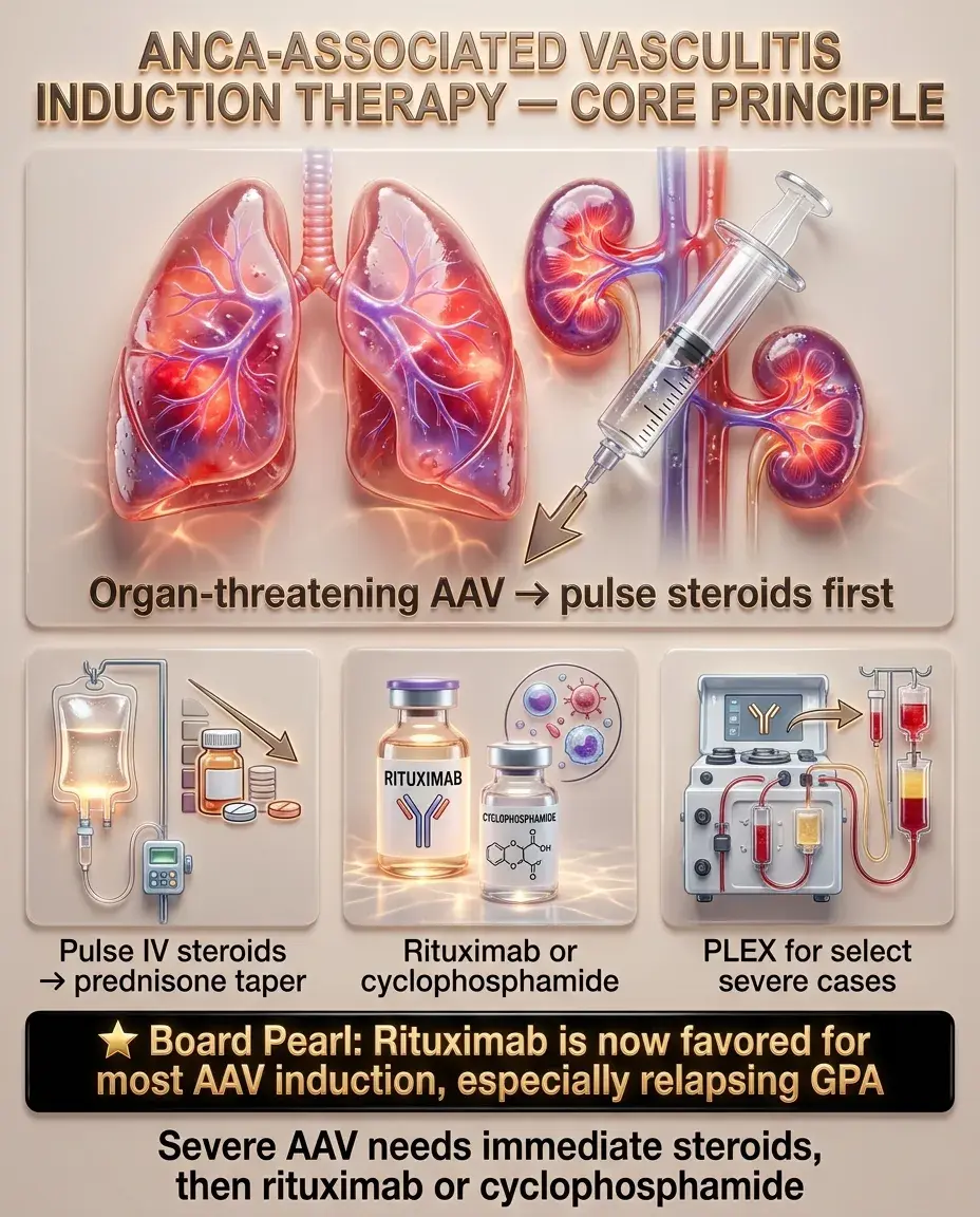
Induction (severe/organ-threatening disease):

— Rituximab preferred for: relapsing disease, PR3-ANCA positivity, fertility concerns, younger patients

— Cyclophosphamide preferred when: rituximab is unavailable, or severe alveolar hemorrhage/refractory disease

Next best step: Organ-threatening AAV → start pulse-dose IV steroids immediately → add rituximab or cyclophosphamide within 1–2 days.

Board pearl: Rituximab has become the favored induction agent for most AAV scenarios, especially relapsing GPA.

Glucocorticoids: pulse IV methylprednisolone (500–1000 mg/day × 3 days) followed by oral prednisone with taper over 3–6 months
Rituximab (anti-CD20) OR cyclophosphamide — both are equivalent for remission induction in GPA and MPA
Plasma exchange (PLEX): consider in severe RPGN (Cr >5.7 mg/dL) or DAH with respiratory failure — evidence is evolving; PEXIVAS trial showed no mortality benefit, but still used in life-threatening cases
Solid White Background
Maintenance Therapy and Treatment Algorithm

Once remission is achieved (typically 3–6 months), transition to maintenance:

Duration of maintenance:

Glucocorticoid taper:

Key distinction: Maintenance agent is chosen based on induction agent, ANCA type, relapse history, and organ involvement — not a one-size-fits-all approach.

Rituximab (preferred): scheduled infusions every 6 months for ≥2 years
Azathioprine: alternative if rituximab is unavailable/contraindicated
Methotrexate: option for limited/non-renal GPA maintenance
Mycophenolate mofetil: second-line; higher relapse rate vs azathioprine in some studies
Minimum 24 months after remission
PR3-ANCA/GPA: higher relapse risk → may need prolonged maintenance
MPO-ANCA/MPA: lower relapse risk → shorter maintenance may suffice
Rapid taper is essential — prolonged steroids ↑ infection risk
Target prednisone discontinuation by 6 months if possible (per reduced-dose steroid protocols showing equivalent efficacy with fewer adverse events)
Solid White Background
Management of EGPA

EGPA treatment differs from GPA/MPA because of its eosinophil-driven pathophysiology:

Non-severe EGPA (no cardiac, renal, GI, or CNS involvement):

Severe EGPA (cardiac, renal, or GI involvement):

Cardiac-specific management:

Board pearl: Mepolizumab is the targeted biologic approved for EGPA — know this for Step 2. It targets IL-5, reducing eosinophil activation.

Key distinction: ANCA-positive EGPA behaves more like classic vasculitis (GN, neuropathy); ANCA-negative EGPA has more cardiac/eosinophilic organ damage.

Glucocorticoids alone may suffice for initial control
Steroid-sparing: azathioprine or methotrexate
Mepolizumab (anti-IL-5): FDA-approved for relapsing/refractory EGPA; reduces eosinophil-mediated damage and allows glucocorticoid dose reduction
Pulse IV steroids + cyclophosphamide
Rituximab: emerging data supporting efficacy in ANCA-positive EGPA
Eosinophilic myocarditis: high-dose steroids + cyclophosphamide
Monitor troponin, BNP, echocardiography
Cardiac MRI for early detection of myocardial involvement
Solid White Background
Special Populations — Pregnancy

AAV in pregnancy:

Medication safety:

Monitoring:

Next best step: Woman of childbearing age with AAV → switch to azathioprine maintenance and ensure ≥3-month washout of MTX/MMF before conception.

Ideally achieve remission ≥6 months before conception
Active vasculitis in pregnancy carries high risk: preeclampsia, IUGR, preterm birth, maternal renal failure
Azathioprine: safe in pregnancy (first-line maintenance during gestation)
Glucocorticoids: generally safe; use lowest effective dose; prednisone preferred (metabolized by placental 11β-HSD)
Rituximab: limited data; generally held before conception (washout ≥6 months); neonatal B-cell depletion reported
Cyclophosphamide: ABSOLUTELY CONTRAINDICATED — teratogenic in first trimester, gonadotoxic
Methotrexate: CONTRAINDICATED — abortifacient, teratogenic; discontinue ≥3 months before conception
Mycophenolate: CONTRAINDICATED — teratogenic
Monthly Cr, urinalysis, CBC
Multidisciplinary: rheumatology + maternal-fetal medicine + nephrology
Solid White Background
Special Populations — Elderly and Renal Impairment

Elderly patients:

Renal impairment:

Board pearl: Infection is the #1 cause of death in the first year after AAV diagnosis — driven by immunosuppression. Prophylaxis and judicious dosing are critical.

MPA is more common in older adults; GPA peaks slightly younger
↑ Infection risk with immunosuppression — leading cause of early mortality in treated AAV
Reduce cyclophosphamide dose if used (age-adjusted dosing)
Rituximab preferred over cyclophosphamide due to lower infection burden
PJP prophylaxis with TMP-SMX is essential when on cyclophosphamide or rituximab
GFR at diagnosis is the strongest predictor of long-term renal outcome
Dialysis-dependent at presentation: still treat with immunosuppression — ~30–50% recover enough to discontinue dialysis
Azathioprine dose adjustment required for renal impairment
Avoid nephrotoxins; monitor drug levels
Cyclophosphamide clearance is reduced in renal failure → dose-adjust to avoid severe myelosuppression
Solid White Background
Complications and When to Escalate Care

Disease-related complications:

Treatment-related complications:

When to escalate:

Next best step: Rising Cr + active sediment in a known AAV patient on maintenance → suspect relapse → recheck ANCA, urinalysis, consider re-biopsy → re-induce.

RPGN → end-stage renal disease: ~20–25% of AAV patients progress to ESRD within 5 years
Diffuse alveolar hemorrhage → respiratory failure → mechanical ventilation
Subglottic stenosis (GPA): may persist despite systemic disease control → requires ENT evaluation, possible dilation/stenting
Eosinophilic myocarditis (EGPA) → cardiogenic shock
Peripheral neuropathy: mononeuritis multiplex may cause foot/wrist drop
Cyclophosphamide: hemorrhagic cystitis (prevent with MESNA + hydration), bladder cancer, infertility, myelosuppression
Rituximab: hypogammaglobulinemia → recurrent infections; check IgG levels before each infusion
Glucocorticoids: osteoporosis, diabetes, infection, AVN
Refractory disease (no response to induction at 4 weeks) → switch rituximab ↔ cyclophosphamide; consider PLEX
Relapse on maintenance → re-induce with rituximab
Solid White Background
Emergencies in ANCA-Associated Vasculitis

Diffuse alveolar hemorrhage (DAH):

Rapidly progressive glomerulonephritis:

Orbital inflammation (GPA):

Board pearl: In DAH, hemoptysis may be absent — a dropping hemoglobin with new bilateral ground-glass opacities should trigger immediate bronchoscopy. Do NOT wait for hemoptysis to act.

Next best step: Suspected pulmonary-renal syndrome → do not delay treatment for ANCA results — start pulse steroids immediately.

Hemoptysis (may be absent!), bilateral ground-glass opacities, ↓ Hgb disproportionate to visible blood loss
BAL shows progressively bloodier returns (diagnostic)
Treatment: pulse IV methylprednisolone + cyclophosphamide or rituximab + consider PLEX
May need intubation and mechanical ventilation
Creatinine doubling over days-weeks + active sediment
Emergency renal biopsy if patient is stable enough
Start immunosuppression empirically while awaiting biopsy/ANCA results if clinical suspicion is high
Proptosis, vision loss → urgent ophthalmology consult + high-dose steroids
Solid White Background
Key Differentials — Pulmonary-Renal Syndrome

Pulmonary-renal syndrome = DAH + GN; differential includes:

— Linear IgG deposits on IF (vs pauci-immune in AAV)

— Anti-GBM antibodies positive

— Young male smoker, rapid course

— Can coexist with AAV ("double-positive") in ~30% of anti-GBM cases

— "Full-house" IF pattern, ↓ complement (C3, C4), ↑ anti-dsDNA

— Other lupus features: malar rash, serositis, cytopenias

— IgA deposits on biopsy; palpable purpura, arthralgias, abdominal pain

— Children > adults

— ↓ complement, ↑ RF, HCV association

— Skin purpura, arthralgias, GN

Key distinction: Normal complement + pauci-immune GN + positive ANCA = AAV. Low complement suggests lupus or cryoglobulinemic vasculitis. Linear IF = anti-GBM.

Anti-GBM disease (Goodpasture syndrome):
SLE nephritis:
IgA vasculitis (HSP):
Cryoglobulinemic vasculitis:
Solid White Background
Key Differentials — GPA vs Other Granulomatous Diseases

GPA vs Sarcoidosis:

GPA vs Relapsing polychondritis:

GPA vs Cocaine-induced midline destructive lesion:

Board pearl: Cocaine-induced midline destruction mimics GPA. The ANCA is typically directed against human neutrophil elastase (HNE) — NOT PR3 or MPO. Always check antigen specificity.

Both cause granulomas, upper/lower respiratory involvement
Sarcoidosis: non-caseating granulomas, bilateral hilar lymphadenopathy, ↑ ACE, ↑ Ca²⁺, non-necrotizing
GPA: necrotizing granulomatous vasculitis, cavitary nodules, ANCA positive, destructive sinusitis
Both can cause saddle-nose deformity
Relapsing polychondritis: auricular chondritis (ear cartilage inflammation sparing lobe), tracheal chondritis, ANCA negative
Cocaine causes nasal septum perforation and positive p-ANCA (anti-human neutrophil elastase, NOT anti-MPO)
Biopsy: necrosis without true vasculitis
Urine drug screen is essential in young patients with destructive nasal lesions
Solid White Background
Preventive Care and Screening

Infection prophylaxis (critical during immunosuppression):

— Also has the bonus of reducing GPA relapse in some studies

— Alternative: dapsone or atovaquone

Vaccinations:

Bone health:

Cyclophosphamide-specific:

PJP prophylaxis: TMP-SMX (preferred) for all patients on cyclophosphamide, rituximab, or high-dose steroids
Hepatitis B screening: before rituximab → reactivation risk
TB screening: before immunosuppression
Update all vaccines before starting immunosuppression
Annual influenza + pneumococcal vaccines
COVID-19 vaccination (antibody response may be blunted post-rituximab)
Avoid live vaccines during immunosuppression
Calcium + vitamin D supplementation
Bisphosphonate if prolonged glucocorticoid use anticipated (≥3 months at prednisone ≥2.5 mg/day)
DEXA scan at baseline
Fertility preservation discussion before treatment (sperm banking, oocyte cryopreservation)
Hemorrhagic cystitis prevention: aggressive hydration + MESNA (IV cyclophosphamide)
Bladder cancer screening: urinalysis annually for life if cumulative dose is high
Solid White Background
Follow-Up and Monitoring

Disease activity monitoring:

Organ-specific surveillance:

Immunosuppression monitoring:

Board pearl: A rising ANCA titer alone — without clinical evidence of disease activity — is NOT an indication to change therapy.

Creatinine and urinalysis with microscopy at every visit (monthly during induction, every 1–3 months on maintenance)
CRP/ESR: helpful trend markers but non-specific
ANCA titers: rising ANCA (especially PR3) may precede clinical relapse, but treatment decisions should be based on clinical disease activity, NOT ANCA titer alone
CBC: monitor for myelosuppression from cyclophosphamide/azathioprine
Renal: GFR trend, proteinuria quantification
Pulmonary: PFTs, chest CT if new symptoms
ENT: regular evaluation in GPA for subglottic stenosis
Cardiac: serial echocardiography in EGPA
Neuropathy: nerve conduction studies if symptoms progress
Rituximab: check IgG levels, CD19/CD20 B-cell counts before each infusion
Azathioprine: TPMT genotype/phenotype before starting → identifies patients at risk for severe myelosuppression
Solid White Background
Ethical, Legal, and Patient Safety Considerations

Board pearl: Always document a fertility preservation discussion before initiating cyclophosphamide — this is both a clinical best practice and a medicolegal standard.

Informed consent for immunosuppression: patients must understand risks of serious infection, malignancy (cyclophosphamide → bladder cancer), and infertility
Fertility counseling: cyclophosphamide is gonadotoxic — discuss fertility preservation before starting, especially in women <35 and men of reproductive age
Shared decision-making: rituximab vs cyclophosphamide — discuss efficacy equivalence, side-effect profiles, insurance coverage, and infusion logistics
Medication adherence: non-adherence to maintenance therapy is a major cause of relapse — structured follow-up and patient education are essential
Transition of care: patients on long-term immunosuppression must have clear documentation of their regimen, prophylaxis, and monitoring plan when transitioning between providers
End-of-life considerations: in elderly patients with advanced CKD from AAV, goals-of-care discussions regarding dialysis and aggressive immunosuppression are appropriate
Solid White Background
High-Yield Associations and Rapid-Fire Facts
PR3-ANCA → GPA; MPO-ANCA → MPA/EGPA
Saddle-nose + sinusitis + cavitary lung nodules + hematuria → GPA
Asthma + eosinophilia + mononeuritis multiplex → EGPA
Pauci-immune crescentic GN = hallmark renal lesion of all AAV
Normal complement levels → distinguishes AAV from lupus/cryoglobulinemic vasculitis
DAH without hemoptysis → dropping Hgb + bilateral GGO → bronchoscopy
Subglottic stenosis may persist despite systemic remission → local therapy needed
TMP-SMX prophylaxis has dual benefit: prevents PJP + may reduce GPA relapse
Cyclophosphamide cumulative dose correlates with bladder cancer risk
EGPA cardiac involvement = #1 cause of death
Rituximab superior for relapsing GPA
ANCA-negative does not exclude AAV (especially EGPA)
Anti-GBM + ANCA double-positive → treat as anti-GBM disease (more aggressive course)
TPMT testing before azathioprine prevents life-threatening myelosuppression
Solid White Background
Board Question Stem Patterns
50M, bloody nasal discharge, saddle-nose, cavitary lung nodules, Cr 3.2, RBC casts → GPA → order PR3-ANCA + renal biopsy → pulse steroids + rituximab
65F, hemoptysis, bilateral GGO, Cr rising rapidly, ANCA positive (MPO) → MPA with DAH + RPGN → pulse steroids + cyclophosphamide or rituximab
38F, refractory asthma, eosinophils 4,500/μL, mononeuritis multiplex, new cardiac dysfunction → EGPA → echocardiography + cardiac MRI → pulse steroids + cyclophosphamide
Patient with GPA in remission, rising PR3-ANCA but no symptoms → Monitor closely but do NOT change therapy based on ANCA alone
Young man, nasal septal perforation, p-ANCA positive, anti-MPO negative → Cocaine-induced midline destruction → urine drug screen
AAV patient on cyclophosphamide, new cough/fever/hypoxia, ground-glass opacities → PJP pneumonia → start TMP-SMX empirically → BAL
EGPA patient relapsing on steroids → Add mepolizumab
Dialysis-dependent AAV patient → Still treat with immunosuppression — renal recovery is possible
AAV with Cr 6.0, requiring dialysis → Consider PLEX in addition to standard induction
Solid White Background
One-Line Recap
ANCA-associated vasculitides (GPA, MPA, EGPA) are pauci-immune, small-vessel necrotizing vasculitides distinguished by ANCA specificity (PR3 → GPA; MPO → MPA/EGPA) and clinical phenotype (GPA: upper/lower airway granulomas + GN; MPA: GN + DAH without granulomas; EGPA: asthma + eosinophilia + cardiac involvement), diagnosed by ANCA serology plus tissue biopsy showing pauci-immune crescentic GN or granulomatous vasculitis, treated with induction (pulse steroids + rituximab or cyclophosphamide) followed by maintenance (rituximab or azathioprine for ≥24 months), with critical attention to infection prophylaxis (TMP-SMX for PJP), fertility counseling before cyclophosphamide, and recognition that rising ANCA alone does not mandate treatment escalation — clinical disease activity drives management decisions.
Solid White Background
Previous Item
Next Item
bottom of page