top of page

Gastrointestinal

Peptic ulcer disease: H. pylori testing and treatment

Clinical Overview and When to Suspect Peptic Ulcer Disease (PUD)

PUD refers to mucosal defects in the stomach or duodenum that extend through the muscularis mucosae, most commonly caused by Helicobacter pylori infection or NSAID use.

— Gnawing/burning epigastric pain, often nocturnal

— Pain improved (duodenal) or worsened (gastric) by eating

— History of NSAID/aspirin use, smoking, or prior H. pylori infection

— Nausea, early satiety, bloating

Board pearl: All patients with documented PUD must be tested for H. pylori — eradication heals ulcers and dramatically reduces recurrence from ~80% to <5%.

Classic patient: middle-aged adult with recurrent epigastric pain, often related to meals
Suspect PUD when:
Duodenal ulcers (DU): ~95% H. pylori-associated; 4× more common than gastric ulcers
Gastric ulcers (GU): ~70% H. pylori; NSAID use is a major contributor
Solid White Background
Presentation Patterns and Key History Findings

Duodenal ulcer pattern:

Gastric ulcer pattern:

Critical history elements:

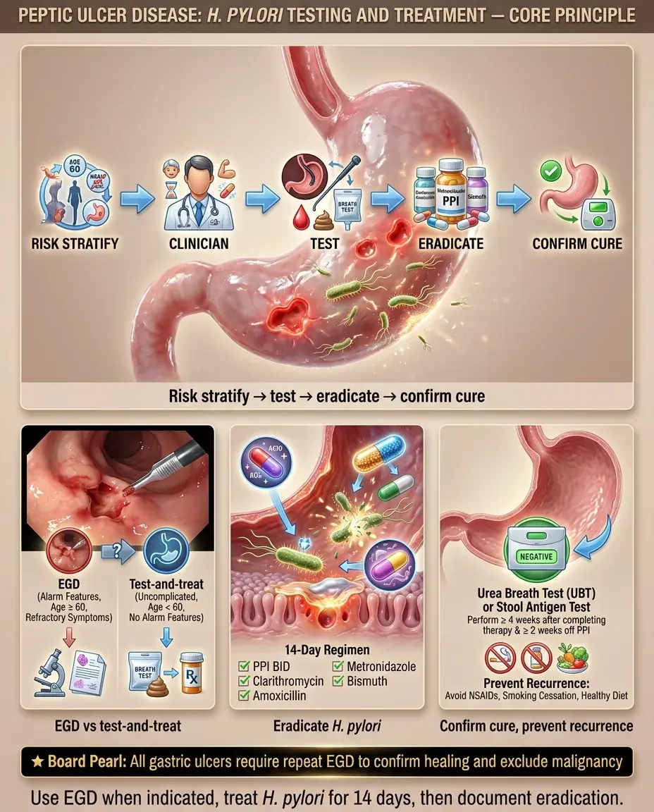
Next best step: Ask about alarm features (see chunk 4) in every patient with dyspepsia — they determine whether to proceed directly to endoscopy vs noninvasive H. pylori testing.

Epigastric pain 2–5 hours after meals and at night ("hunger pain")
Pain relieved by food or antacids
Rarely malignant
Epigastric pain worsened by or occurring with meals
Associated weight loss, nausea, early satiety
Must rule out malignancy (biopsy required at endoscopy)
NSAID/aspirin/corticosteroid use (especially combined)
Anticoagulant/antiplatelet agents → ↑ bleeding risk
Smoking → delays ulcer healing, ↑ recurrence
Alcohol use → mucosal irritation
Prior PUD episodes or H. pylori treatment
Family history of gastric cancer
Solid White Background
Physical Exam and Signs of Complications

Uncomplicated PUD:

Signs suggesting complications:

Board pearl: Succussion splash (sloshing sound with gentle rocking) suggests retained gastric contents → think GOO from chronic PUD or malignancy.

Exam is often unremarkable
Mild epigastric tenderness without peritoneal signs
GI bleeding: hematemesis, melena, coffee-ground emesis, tachycardia, orthostatic hypotension, pallor
Perforation: sudden severe epigastric pain → diffuse peritonitis, rigid "board-like" abdomen, absent bowel sounds, Mackenzie sign (referred shoulder pain from diaphragmatic irritation)
Gastric outlet obstruction (GOO): projectile nonbilious vomiting, succussion splash, visible gastric peristalsis, hypochloremic hypokalemic metabolic alkalosis
Penetration (posterior DU → pancreas): pain radiating to back, ↑ lipase
Solid White Background
Diagnostic Workup — Indications for EGD vs Noninvasive Testing

Alarm features mandating esophagogastroduodenoscopy (EGD):

Approach without alarm features (age <60):

If EGD performed:

Key distinction: Duodenal ulcers rarely need follow-up EGD; gastric ulcers always need repeat EGD to confirm healing and exclude cancer.

Age ≥60 with new-onset dyspepsia
Unintentional weight loss
Progressive dysphagia/odynophagia
GI bleeding (overt or occult)/iron-deficiency anemia
Persistent vomiting
Palpable abdominal mass
Family history of upper GI malignancy
"Test-and-treat" strategy for H. pylori is first-line
Noninvasive testing → treat if positive → reassess
DU: biopsy not routinely needed (rarely malignant); biopsy for H. pylori
GU: always biopsy ulcer edges (rule out malignancy) + biopsy for H. pylori
Repeat EGD in 8–12 weeks for GU to document healing
Solid White Background
H. pylori Testing — Noninvasive and Invasive Methods

Noninvasive tests:

Invasive tests (during EGD):

Critical pre-test requirements:

Board pearl: Serology is the only test unaffected by PPI/antibiotic use but cannot be used for test-of-cure because IgG remains positive long after eradication.

Urea breath test (UBT): sensitivity/specificity >95%; best for initial diagnosis and confirming eradication
Stool antigen test (SAT): sensitivity/specificity >95%; equivalent to UBT; preferred if UBT unavailable
Serology (IgG): sensitivity ~85%; does NOT distinguish active vs past infection → cannot confirm eradication; avoid for test-of-cure
Rapid urease test (CLO test): biopsy-based; sensitivity ~90–95%; result in hours
Histology: gold standard; also assesses for intestinal metaplasia/dysplasia
Culture: low sensitivity; used when antibiotic resistance testing needed
Stop PPIs ≥2 weeks before testing
Stop antibiotics and bismuth ≥4 weeks before testing
Reason: these suppress organism load → false-negative results on all tests except serology
Solid White Background
First-Line H. pylori Eradication — Bismuth Quadruple and Clarithromycin Triple Therapy

First-line regimen selection depends on local clarithromycin resistance (<15% → triple OK; ≥15% or unknown → quadruple preferred):

Bismuth quadruple therapy (preferred if resistance unknown):

Clarithromycin-based triple therapy (only if local resistance <15% AND no prior macrolide exposure):

Key principles:

Next best step: Before prescribing, ask about penicillin allergy and prior macrolide exposure — these change the regimen.

PPI (standard dose BID) + bismuth subsalicylate + metronidazole + tetracycline × 14 days
Avoids clarithromycin resistance issue
PPI (BID) + clarithromycin + amoxicillin × 14 days
Alternative: PPI + clarithromycin + metronidazole (if penicillin allergy)
14-day courses are superior to 7- or 10-day regimens
Always use PPI BID, not once daily
Smoking cessation improves eradication rates
Solid White Background
Alternative and Salvage H. pylori Regimens

Concomitant therapy (increasingly used first-line):

Levofloxacin-based triple therapy (salvage/second-line):

Rifabutin-based triple therapy (third-line/refractory):

General salvage principles:

Board pearl: After first-line failure, switch to a regimen with completely different antibiotics — do not repeat the same class.

PPI (BID) + clarithromycin + amoxicillin + metronidazole × 14 days
Overcomes single-antibiotic resistance by using all three agents simultaneously
PPI (BID) + levofloxacin + amoxicillin × 14 days
Reserve for treatment failure; fluoroquinolone resistance is rising
PPI (BID) + rifabutin + amoxicillin × 14 days
Risk of myelotoxicity; used when ≥2 prior regimens fail
Never re-use clarithromycin or levofloxacin after failure with those agents
Consider culture with susceptibility testing after ≥2 failed regimens
Ensure compliance — nonadherence is a major cause of treatment failure
Solid White Background
Treatment Algorithm and Acid Suppression in PUD

PUD management overview:

1. Test for H. pylori → eradicate if positive

2. Discontinue NSAIDs if possible

3. PPI therapy to heal ulcer:

— DU: PPI × 4–8 weeks

— GU: PPI × 8–12 weeks

4. Confirm H. pylori eradication (test-of-cure)

5. GU: repeat EGD at 8–12 weeks to document healing

If H. pylori-negative and NSAID-negative:

PPI dosing for ulcer healing:

Key distinction: NSAID-related ulcers require NSAID cessation as the primary intervention; H. pylori eradication alone does not heal NSAID-induced ulcers if the drug is continued.

Exclude other causes: Zollinger-Ellison syndrome (gastrinoma), Crohn disease, CMV (immunosuppressed)
Check fasting serum gastrin level if refractory/multiple ulcers
Omeprazole 20 mg BID (for eradication), then 20 mg daily (for healing)
H2 blockers are inferior to PPIs for ulcer healing
Solid White Background
Special Populations — Pregnancy, Pediatrics, Elderly

Pregnancy:

Pediatric considerations:

Elderly:

Board pearl: In pregnancy, defer H. pylori eradication if possible; bismuth and tetracycline are absolutely contraindicated.

PPIs (omeprazole, pantoprazole) are generally safe (FDA category B/C)
Avoid bismuth (category D), metronidazole (avoid first trimester), tetracycline (contraindicated)
Amoxicillin + clarithromycin + PPI can be considered if eradication is necessary
Often defer eradication until postpartum unless clinically urgent
H. pylori testing recommended only for symptomatic children (not routine screening)
EGD-based diagnosis preferred over noninvasive testing in children
Treatment: PPI + amoxicillin + clarithromycin (or metronidazole) × 14 days
↑ Risk of PUD complications (bleeding, perforation) due to NSAID polypharmacy, anticoagulants
Lower threshold for EGD (alarm features threshold: age ≥60)
Monitor renal function — adjust medications accordingly
Long-term PPI use concerns: ↑ risk of C. difficile, osteoporotic fractures, hypomagnesemia, B12 deficiency
Solid White Background
NSAID-Related PUD and Gastroprotection Strategies

Risk factors for NSAID-induced PUD:

Gastroprotection strategies:

Aspirin users:

Key distinction: H. pylori and NSAIDs are independent, synergistic risk factors. Eradicating H. pylori reduces but does not eliminate NSAID ulcer risk — PPI co-therapy is still needed in high-risk patients on chronic NSAIDs.

Next best step: Before starting long-term NSAID therapy in a patient with PUD history → test for H. pylori, eradicate if positive, AND add PPI.

Age >65, prior PUD/GI bleed, concurrent anticoagulant/antiplatelet/corticosteroid use
High-dose or multiple NSAIDs, H. pylori co-infection
If NSAID is essential and patient has risk factors → co-prescribe PPI
Substitute COX-2 selective inhibitor (celecoxib) + PPI for highest-risk patients
Test and treat H. pylori before chronic NSAID use
Low-dose aspirin + prior GI bleed → add PPI indefinitely
Dual antiplatelet therapy (DAPT) → PPI co-therapy recommended
Solid White Background
Complications — Upper GI Bleeding From PUD

PUD is the most common cause of upper GI bleeding (UGIB).

Initial management:

EGD timing:

Forrest classification (endoscopic findings guiding rebleeding risk):

Board pearl: IV PPI infusion is started empirically before EGD in suspected bleeding PUD — it stabilizes clots and reduces the need for endoscopic intervention.

ABCs, two large-bore IV lines, fluid resuscitation
Type and crossmatch; transfuse pRBCs if Hgb <7 g/dL (threshold ↑ to <8 in active CAD)
IV PPI bolus (80 mg) then continuous infusion (8 mg/hr) before EGD
Prokinetic (IV erythromycin 250 mg) 30–60 min pre-EGD to clear gastric blood → improves visualization
Within 24 hours for most UGIB
Emergent (<12 hours) if hemodynamically unstable despite resuscitation
Active spurting (Ia) or oozing (Ib) → endoscopic therapy
Visible vessel (IIa) → endoscopic therapy
Adherent clot (IIb) → attempt removal → treat underlying lesion
Flat pigmented spot (IIc) or clean base (III) → low rebleeding risk → no endoscopic therapy needed
Solid White Background
Complications — Perforation and Gastric Outlet Obstruction

Perforation:

Gastric outlet obstruction (GOO):

Board pearl: Anterior DU → perforation; posterior DU → hemorrhage (gastroduodenal artery erosion).

Most common with anterior duodenal ulcers
Sudden onset severe epigastric pain → diffuse peritonitis
Upright chest X-ray/abdominal X-ray: free air under diaphragm (pneumoperitoneum) — sensitivity ~80%
If X-ray negative but high suspicion → CT abdomen/pelvis with oral contrast (sensitivity >95%)
Next best step: NPO, IV fluids, IV antibiotics, emergent surgical consultation → typically Graham patch (omental patch repair)
Posterior DU perforation can erode into gastroduodenal artery → massive hemorrhage
Chronic scarring from recurrent ulcers → pyloric stenosis
Presents with early satiety, nonbilious vomiting, weight loss
Metabolic derangement: hypochloremic, hypokalemic metabolic alkalosis (loss of HCl)
Diagnosis: EGD (preferred — also therapeutic) or barium swallow showing narrowing
Treatment: endoscopic balloon dilation; surgery if refractory
Solid White Background
Key Differentials — Dyspepsia and Epigastric Pain

Functional dyspepsia:

Gastric cancer:

Zollinger-Ellison syndrome (ZES):

Key distinction: Isolated gastric ulcer that fails to heal after 12 weeks of PPI + H. pylori eradication → biopsy to exclude malignancy or consider ZES.

Chronic epigastric pain/burning without ulcer on EGD
Rome IV criteria: ≥3 months, onset ≥6 months prior
Management: test-and-treat H. pylori → PPI trial → tricyclic antidepressant
Alarm features: weight loss, early satiety, dysphagia, anemia, palpable mass
EGD with biopsy of all gastric ulcers is mandatory
Risk factors: H. pylori (especially intestinal-type), smoking, salt-preserved foods, family history
Multiple/refractory ulcers, ulcers in unusual locations (distal duodenum/jejunum)
Profuse diarrhea (acid hypersecretion)
Fasting gastrin >1000 pg/mL with gastric pH <2 → diagnostic
Secretin stimulation test if gastrin equivocal
Associated with MEN1 (pituitary, parathyroid, pancreas)
Solid White Background
Distinguishing H. pylori-Related vs NSAID-Related PUD

H. pylori-related PUD:

NSAID-related PUD:

Both present:

Board pearl: NSAID ulcers are commonly asymptomatic until complications occur — this is why gastroprophylaxis with PPI is recommended in high-risk patients on chronic NSAIDs.

Multiple or recurrent ulcers
Duodenal > gastric ulcers
Chronic active gastritis on biopsy
Eradication cures disease; ulcer recurrence rate drops from ~80% → <5%
Associated with gastric MALT lymphoma and gastric adenocarcinoma
Gastric > duodenal ulcers
Often painless — may present with bleeding as first symptom (especially elderly)
Mucosal damage via COX-1 inhibition → ↓ prostaglandins → ↓ mucosal protection
Removal of offending agent is critical to healing
H. pylori testing still needed — dual etiology possible
Synergistic risk; eradicate H. pylori AND stop NSAIDs if possible
Solid White Background
Test-of-Cure and Post-Treatment Follow-Up

Confirming H. pylori eradication:

Follow-up EGD:

If eradication fails:

Next best step: After completing H. pylori therapy, wait ≥4 weeks off antibiotics and ≥2 weeks off PPIs before performing UBT or stool antigen for test-of-cure.

Test-of-cure is recommended for ALL patients treated for H. pylori
Use UBT or stool antigen test (NOT serology — remains positive)
Timing: ≥4 weeks after completing antibiotics AND ≥2 weeks after stopping PPI
Rationale: ensures eradication, prevents ulcer recurrence, reduces cancer risk
Gastric ulcers: repeat EGD at 8–12 weeks to confirm healing and rebiopsy if not healed
Duodenal ulcers: no routine repeat EGD unless symptoms persist
Use a different regimen (see chunk 7)
Confirm medication compliance
After ≥2 failures → culture with antibiotic susceptibility testing
Solid White Background
Screening, Prevention, and Long-Term Monitoring

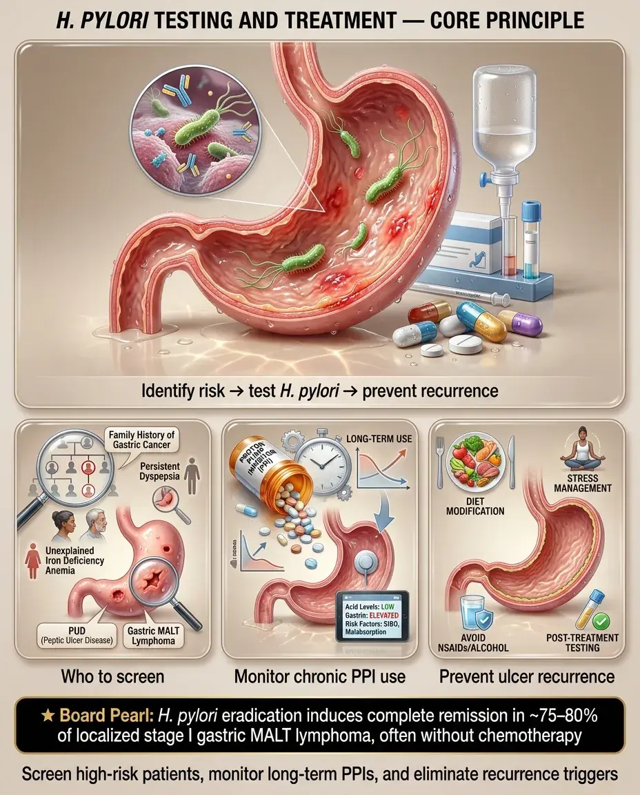
H. pylori screening indications (even without active PUD):

Long-term PPI monitoring (if ongoing PPI needed):

Prevention of PUD recurrence:

Board pearl: H. pylori eradication in gastric MALT lymphoma achieves complete remission in ~75–80% of localized (stage I) cases — chemotherapy may not be needed.

Active or history of PUD
Gastric MALT lymphoma
Early gastric cancer (after endoscopic resection)
First-degree relative with gastric cancer
Unexplained iron deficiency anemia (after other causes excluded)
Chronic idiopathic thrombocytopenic purpura (ITP)
Before long-term NSAID or aspirin therapy
Ca²⁺ and vitamin D supplementation for osteoporosis risk
Monitor Mg²⁺ levels (hypomagnesemia with chronic use)
B12 levels in elderly on long-term therapy
Reassess PPI necessity annually — step down to lowest effective dose or H2 blocker
Eradicate H. pylori
Avoid NSAIDs; use COX-2 inhibitors + PPI if unavoidable
Smoking cessation
Limit alcohol
Solid White Background
Ethical, Legal, and Patient Safety Considerations

Informed consent and shared decision-making:

Antibiotic stewardship:

Medication safety:

Patient adherence:

Board pearl: Nonadherence is the most common cause of H. pylori treatment failure — address it before changing antibiotics.

Discuss risks/benefits of EGD, including sedation risks, perforation (~1:10,000), and bleeding
Patients declining EGD → document informed refusal; pursue noninvasive H. pylori testing
Avoid unnecessary clarithromycin use in areas of high resistance
Use susceptibility-guided therapy when available (especially after treatment failures)
Complete 14-day courses to minimize resistance development
Clarithromycin: QT prolongation risk; check for drug interactions (statins, colchicine, warfarin)
Metronidazole: disulfiram-like reaction with alcohol; peripheral neuropathy with prolonged use
Tetracycline: photosensitivity, contraindicated in pregnancy/children <8
PPIs: educate patients on appropriate duration — avoid indefinite use without indication
Quadruple therapy regimens are complex (4 drugs, multiple doses) → pill burden is high
Counsel on importance of completing full course
Side effects (metallic taste, nausea, diarrhea) are common → preemptive counseling improves adherence
Solid White Background
High-Yield Associations and Rapid-Fire Clinical Facts

Board pearl: H. pylori ↑ risk of both gastric adenocarcinoma and MALT lymphoma but is inversely associated with esophageal adenocarcinoma and GERD.

H. pylori → chronic active gastritis → atrophic gastritis → intestinal metaplasia → dysplasia → gastric adenocarcinoma (Correa cascade)
H. pylori is a WHO Class I carcinogen
Duodenal ulcer: anterior → perforation; posterior → bleeding (gastroduodenal artery)
Gastric MALT lymphoma: eradicate H. pylori first → most regress
ZES: fasting gastrin >1000 + pH <2 = diagnostic; secretin stimulation test if equivocal
Serology = only H. pylori test NOT affected by PPI/antibiotics
UBT and stool antigen = best for test-of-cure
NSAID ulcers are more often painless → present with bleeding
Currant jelly sputum = Klebsiella (don't confuse — PUD context: currant jelly not relevant)
Misoprostol (PGE₁ analog) → mucosal protection but limited by diarrhea/uterine contractions; PPI preferred for gastroprotection
Coffee-ground emesis + melena → upper GI source → PUD until proven otherwise
Solid White Background
Board Question Stem Patterns
45M, burning epigastric pain relieved by eating, no alarm features, age <60 → Test-and-treat H. pylori (UBT or stool antigen)
62F, new dyspepsia + 10-lb weight loss → EGD with biopsy (alarm features)
EGD shows gastric ulcer, rapid urease test positive → H. pylori eradication (bismuth quadruple × 14 days) + PPI × 8–12 weeks + repeat EGD to confirm healing
Patient completed triple therapy, wants test-of-cure → Wait ≥4 weeks after antibiotics, ≥2 weeks after PPI → UBT or stool antigen (NOT serology)
Duodenal ulcer on chronic NSAIDs, H. pylori positive → Eradicate H. pylori AND stop NSAID AND PPI to heal ulcer
Sudden severe epigastric pain, rigid abdomen, free air on CXR → Perforated ulcer → IV fluids, antibiotics, emergent surgical repair
Multiple refractory ulcers in unusual location + diarrhea → ZES → fasting gastrin level
GI bleed from DU, visible vessel on EGD → Endoscopic hemostasis (epinephrine + thermal/clips) + IV PPI infusion
First-line H. pylori eradication failed → Switch to different regimen (e.g., bismuth quadruple if triple used first; levofloxacin-based if quadruple failed) — never repeat same antibiotics
Stage I gastric MALT lymphoma, H. pylori positive → Eradicate H. pylori → reassess; ~80% achieve remission
Solid White Background
One-Line Recap
Peptic ulcer disease is most commonly caused by H. pylori infection or NSAIDs, diagnosed by EGD (required when alarm features present) or noninvasive test-and-treat strategy (UBT/stool antigen for age <60 without alarm features), treated with 14-day H. pylori eradication using bismuth quadruple therapy (preferred) or clarithromycin triple therapy (if low resistance), PPI for 4–12 weeks to heal the ulcer, NSAID cessation when possible, with mandatory test-of-cure via UBT or stool antigen ≥4 weeks post-antibiotics, repeat EGD for all gastric ulcers to exclude malignancy, and gastroprotection (PPI ± COX-2 inhibitor) for patients requiring continued NSAID/aspirin therapy who have risk factors for ulcer complications.
Solid White Background
Previous Item
Next Item
bottom of page