top of page

Nervous System

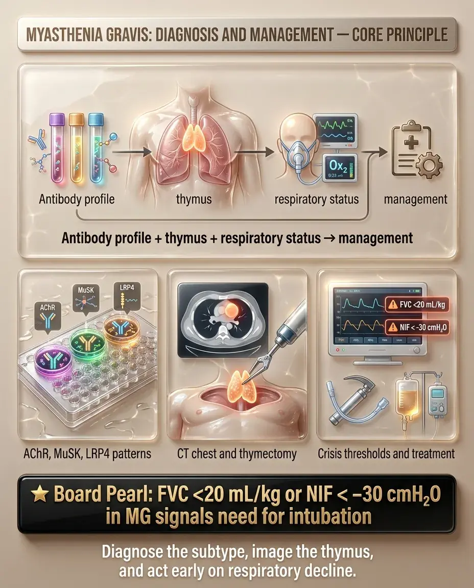
Myasthenia gravis: diagnosis and management

Clinical Overview and When to Suspect Myasthenia Gravis

Myasthenia gravis (MG) is an autoimmune disorder caused by antibodies against the postsynaptic neuromuscular junction (NMJ) — most commonly anti-acetylcholine receptor (AChR) antibodies (~85%) → ↓ functional AChR → fatigable weakness.

— Ptosis and diplopia (ocular involvement is the most common initial presentation)

— Difficulty chewing, swallowing, speaking (bulbar weakness)

— Proximal limb weakness

— Symptoms worse at the end of the day

Board pearl: MG affects the postsynaptic NMJ — distinguishing it from Lambert-Eaton (presynaptic) and botulism (presynaptic) is heavily tested.

Classic patient: young woman (20–30s) or older man (60–70s) with fluctuating, fatigable weakness that worsens with repeated use and improves with rest
Hallmark features:
Suspect MG when weakness is fatigable, fluctuating, painless, and involves ocular/bulbar muscles without sensory loss
Solid White Background
Presentation Patterns and Key History

— Asymmetric ptosis, fluctuating diplopia

— Pupils always spared (unlike CN III palsy from compression)

— Ocular → bulbar → limb/respiratory progression

— Bulbar: dysarthria, dysphagia, nasal voice, jaw fatigue while chewing

— Limb: proximal > distal, arms often > legs

— Respiratory: dyspnea, inability to count to 20 in one breath

Key history clues:

— Symptoms worse with activity, heat, stress, illness, menses

— Associated autoimmune conditions (thyroid disease, RA, SLE)

— Drug-induced exacerbation: aminoglycosides, fluoroquinolones, β-blockers, magnesium, neuromuscular blockers, D-penicillamine

— Thymoma association (10–15%) — ask about chest imaging findings

Next best step: When a patient reports fluctuating diplopia/ptosis with fatigability → test for AChR antibodies.

Ocular MG (~50% present here, ~15% remain purely ocular):
Generalized MG:
Solid White Background
Physical Exam Findings and Bedside Tests

Important negatives:

— Sensation fully intact

— Reflexes normal

— No fasciculations (distinguishes from ALS)

— Pupils normal

Board pearl: The ice pack test is a safe, noninvasive bedside test for MG-related ptosis — improvement supports diagnosis, especially when edrophonium testing is not readily available.

Fatigable ptosis: sustained upgaze for 60 seconds → progressive ptosis
Curtain sign: lifting the more ptotic eyelid → contralateral lid droops further (orbicularis weakness distributes effort)
Ice pack test: apply ice to closed eyelid for 2 minutes → improvement of ptosis (cold inhibits AChE → ↑ ACh at NMJ) — quick bedside screen
Peek sign: ask patient to gently close eyes — lids slowly separate due to orbicularis oculi fatigue
Cogan lid twitch: after sustained downgaze, return to primary gaze → brief upward overshoot of lid
Solid White Background
Diagnostic Workup — Serological Testing

Step 1 — Antibody testing (most important):

— Highly specific (>99%); positive result essentially confirms diagnosis

— Three types: binding, blocking, modulating — binding is most sensitive

— MuSK-MG: prominent bulbar/facial/respiratory weakness, less ocular

— Often younger women; may have tongue/facial atrophy

— Poor response to AChE inhibitors; responds better to rituximab

Step 2 — Confirm with electrophysiology if antibodies negative:

Board pearl: AChR antibodies are the first-line diagnostic test. A positive result in the right clinical context is diagnostic — no further NMJ testing needed.

Anti-AChR antibodies: positive in ~85% of generalized MG, ~50% of ocular MG
If AChR-Ab negative → test anti-MuSK antibodies (~5–8% of MG)
If both negative → anti-LRP4 antibodies (rare); or "seronegative" MG (~5–10%)
Repetitive nerve stimulation (RNS)
Single-fiber EMG (SFEMG)
Solid White Background
Diagnostic Workup — Electrophysiology and Imaging

Repetitive nerve stimulation (RNS):

Single-fiber EMG (SFEMG):

CT chest (all MG patients):

Key distinction: Edrophonium (Tensilon) test is rarely used now due to cardiac risks (bradycardia) and availability of better diagnostics — antibody testing + EMG have replaced it.

Low-frequency (2–3 Hz) stimulation → decremental response (>10% decrement from 1st to 4th/5th potential)
Sensitivity: ~75% generalized MG, ~30–50% ocular MG
This is postsynaptic pattern; contrast with Lambert-Eaton → incremental response at high-frequency stimulation
Most sensitive test for MG (>95% sensitivity)
Shows ↑ jitter (variability in NMJ transmission time)
Used when antibodies and RNS are negative but clinical suspicion remains high
Screen for thymoma — present in 10–15% of MG patients
Thymic hyperplasia in ~65% (especially young-onset MG)
If thymoma found → surgical resection regardless of MG subtype
Solid White Background
First-Line Management — Symptomatic Therapy

Acetylcholinesterase (AChE) inhibitors — first-line symptomatic treatment:

— Inhibits AChE → ↑ ACh at NMJ → improved neuromuscular transmission

— Onset 30 min, duration 3–4 hours

— May suffice as sole therapy in mild/ocular MG

Side effects (cholinergic):

— Muscarinic: diarrhea, abdominal cramps, ↑ salivation, bradycardia, miosis

— Nicotinic (overdose): fasciculations, weakness — can mimic worsening MG (cholinergic crisis)

— Add glycopyrrolate for muscarinic side effects if needed

Important: Pyridostigmine does NOT alter disease course — it is purely symptomatic. Most patients with generalized MG will need immunotherapy.

Board pearl: MuSK-MG responds poorly to pyridostigmine; consider rituximab early in MuSK-positive disease.

Pyridostigmine (Mestinon) 60 mg PO q4–6h; titrate to symptom control
Solid White Background
Immunosuppressive Therapy — Disease Modification

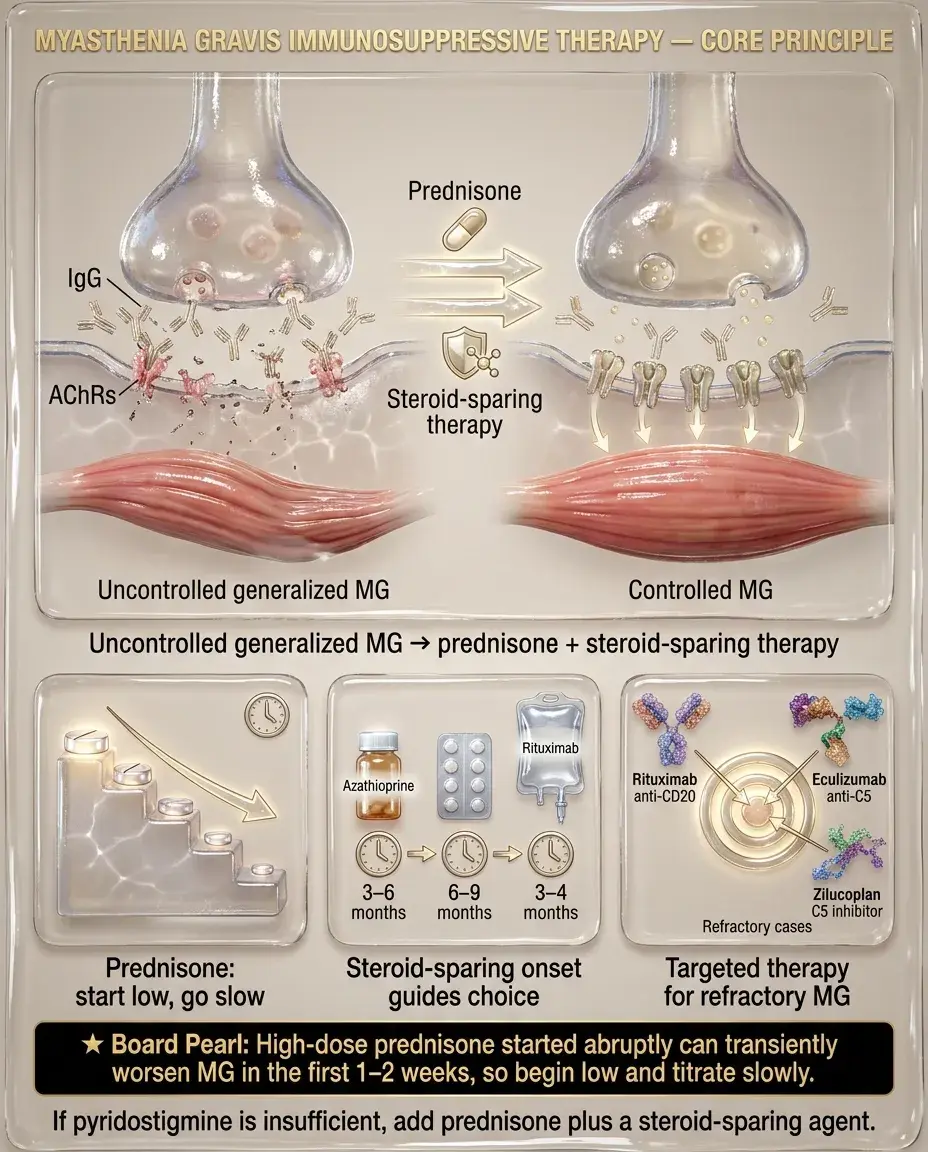
Corticosteroids — first-line immunotherapy for moderate-severe MG:

— Rapid initiation at high dose can paradoxically worsen weakness in first 1–2 weeks ("steroid dip") → start low, go slow, or initiate in-hospital for severe disease

— Once stable, taper to lowest effective dose

Steroid-sparing agents (used with or instead of steroids):

Newer targeted therapy:

Next best step: Generalized MG not controlled by pyridostigmine → add prednisone + steroid-sparing agent.

Prednisone: start low (10–20 mg/day), titrate slowly upward
Azathioprine — most commonly used; onset 6–12 months; check TPMT before starting
Mycophenolate mofetil — alternative; onset 6–12 months
Cyclosporine or tacrolimus — faster onset (weeks), used if azathioprine/MMF intolerable
Rituximab (anti-CD20) — particularly effective in MuSK-MG
Eculizumab (anti-C5 complement inhibitor) — FDA-approved for AChR-Ab+ refractory generalized MG
Efgartigimod (FcRn blocker) — ↓ pathogenic IgG; approved for AChR-Ab+ generalized MG
Solid White Background
Thymectomy — Indications and Role

Thymectomy indications:

Not recommended:

Timing and approach:

Board pearl: Even without thymoma, thymectomy improves outcomes in AChR-Ab+ generalized MG — this is a key Step 2 CK concept from the landmark MGTX randomized trial.

Thymoma — absolute indication regardless of MG severity or antibody status
Generalized AChR-Ab+ MG (age 18–65) without thymoma — MGTX trial showed improved outcomes (↓ prednisone requirement + better symptom control over 3 years)
Consider in refractory ocular MG converting to generalized
MuSK-MG — thymus is typically normal; no proven benefit from thymectomy
Very elderly or significant comorbidities where surgical risk outweighs benefit
Optimize MG control before surgery (IVIG/PLEX pre-op if needed)
Robotic/minimally invasive preferred; ensure complete resection
Benefits may take months to years to fully manifest
Solid White Background
Special Populations — Pregnancy and Pediatric MG

Pregnancy:

Pediatric MG:

— Suspect if onset at birth, family history, seronegative

Board pearl: Never give magnesium sulfate to a pregnant patient with MG — it can precipitate myasthenic crisis.

MG can worsen, improve, or remain stable — unpredictable
Exacerbations most common in 1st trimester and postpartum
Safe medications: pyridostigmine, IVIG, prednisone (low dose)
Avoid: mycophenolate (teratogenic — Category X), methotrexate
Azathioprine: relatively safe but discuss risks/benefits
Magnesium sulfate (for preeclampsia) is CONTRAINDICATED — blocks NMJ → precipitates crisis
Neonatal MG: transient weakness in 10–20% of neonates born to MG mothers (passive transfer of AChR-Ab) → self-resolves in weeks
Juvenile MG: autoimmune, similar to adult; AChR-Ab often positive
Congenital myasthenic syndromes: genetic (not autoimmune) → do not respond to immunotherapy
Solid White Background
Special Populations — Elderly and Comorbidities

Elderly-onset MG (>60 years):

Renal impairment:

Hepatic impairment:

Drug interactions — medications that worsen MG:

Next best step: Review medication list in every MG patient — drug-induced exacerbation is a preventable trigger.

Male predominance; higher association with thymoma
More likely to present with bulbar/generalized weakness
AChR antibodies often positive at high titers
Comorbidities (COPD, cardiac disease) increase risk of respiratory crisis
Steroid side effects more pronounced: osteoporosis, hyperglycemia, cataracts → emphasize steroid-sparing agents early
Pyridostigmine partially renally excreted → ↓ dose in CKD
Azathioprine dose adjustment may be needed; monitor CBC closely
Mycophenolate, azathioprine hepatotoxic → monitor LFTs
Cyclosporine: hepato-/nephrotoxic — avoid if possible
Aminoglycosides, fluoroquinolones, macrolides
β-blockers, calcium channel blockers (verapamil)
Neuromuscular blocking agents (prolonged paralysis post-anesthesia)
Magnesium, D-penicillamine, checkpoint inhibitors
Solid White Background
Myasthenic Crisis — Recognition and Emergency Management

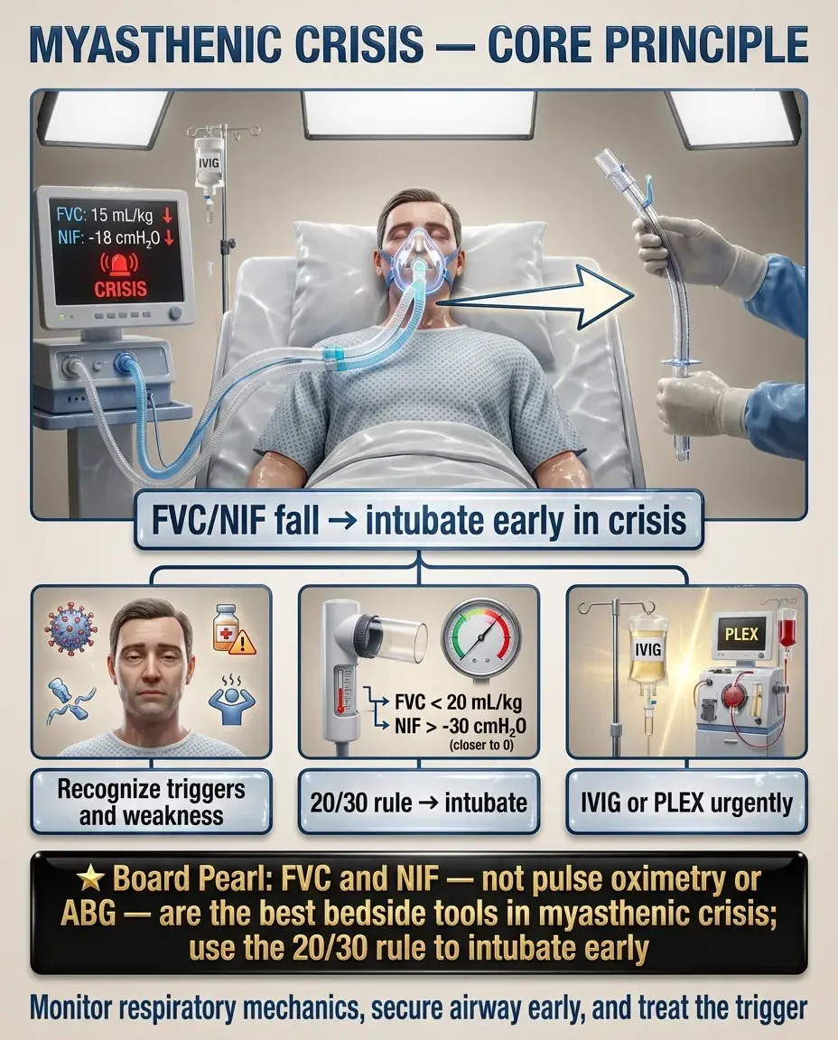
Myasthenic crisis: life-threatening exacerbation with respiratory failure requiring mechanical ventilation — occurs in ~15–20% of MG patients.

— FVC <20 mL/kg or NIF weaker than −30 cmH₂O → intubate (do not wait for ABG deterioration)

Acute treatment:

— Equally effective; IVIG preferred if IV access is difficult

— PLEX faster onset (days); IVIG easier to administer

Board pearl: FVC and NIF — not pulse oximetry or ABG — are the best bedside tools to monitor respiratory function in MG crisis. The "20/30 rule": FVC <20 mL/kg or NIF weaker than −30 cmH₂O → intubate.

Triggers: infection (most common), surgery, medication changes, medication non-adherence, offending drugs
Monitor: forced vital capacity (FVC) and negative inspiratory force (NIF)
IVIG (0.4 g/kg/day × 5 days) or plasma exchange (PLEX, 5 exchanges over 10–14 days)
Continue or increase immunosuppression
Treat underlying trigger (antibiotics for infection, etc.)
Solid White Background
Cholinergic Crisis vs Myasthenic Crisis

Key distinction tested on boards:

— Pupils normal, dry secretions or normal

— Treatment: IVIG/PLEX + ventilatory support

— SLUDGE signs: Salivation, Lacrimation, Urination, Diarrhea, GI cramping, Emesis

— Bradycardia, miosis, fasciculations, excessive bronchial secretions

— Treatment: stop pyridostigmine, atropine for muscarinic effects, ventilatory support

Both present with worsening weakness — differentiation:

— Muscarinic excess → cholinergic crisis

— No muscarinic signs → myasthenic crisis

Board pearl: In a weak MG patient on pyridostigmine with excessive secretions and bradycardia → cholinergic crisis → stop pyridostigmine, give atropine.

Myasthenic crisis: worsening of disease → ↓ ACh effect at NMJ → weakness + respiratory failure
Cholinergic crisis: excessive AChE inhibitor (pyridostigmine overdose) → ↑ ACh → depolarization block + muscarinic overload
In practice, both may coexist → hold AChE inhibitors, support ventilation, reassess
Solid White Background
Key Differentials — Ptosis and Diplopia

Key distinction: Fatigable weakness + pupil-sparing + no sensory loss → think MG. Fixed deficits, pupil involvement, or sensory findings → alternative diagnosis.

CN III palsy (compressive: aneurysm): pupil dilated + ptosis + "down and out" eye → pupil involvement distinguishes from MG
CN III palsy (microvascular: DM/HTN): pupil spared, but weakness is fixed, not fatigable
Horner syndrome: mild ptosis + miosis + anhidrosis → sympathetic disruption; pupil constricted (opposite of CN III)
Thyroid eye disease (Graves): proptosis, lid retraction/lag, restrictive ophthalmopathy — not fatigable
Chronic progressive external ophthalmoplegia (CPEO): mitochondrial; slowly progressive bilateral ptosis + ophthalmoplegia; not fatigable
Miller Fisher syndrome (GBS variant): ophthalmoplegia + ataxia + areflexia; acute onset, anti-GQ1b antibodies
Solid White Background
Key Differentials — Generalized Weakness

— Presynaptic (anti-VGCC antibodies) vs MG (postsynaptic)

— Proximal weakness that IMPROVES with repeated use (opposite of MG)

— Autonomic dysfunction: dry mouth, constipation, erectile dysfunction

— Associated with small cell lung cancer (~60%)

— RNS: incremental response at high frequency (opposite of MG's decremental)

Board pearl: LEMS improves with activity, MG worsens — this distinction is a classic board differentiator. Always screen LEMS patients for small cell lung cancer.

Lambert-Eaton myasthenic syndrome (LEMS):
Botulism: presynaptic; descending paralysis, dilated unreactive pupils, autonomic dysfunction; history of wound/canned food
ALS: upper + lower motor neuron signs; fasciculations, no sensory loss; progressive, not fatigable
Polymyositis/dermatomyositis: proximal weakness, ↑ CK, no ocular involvement, muscle biopsy inflammatory infiltrate
GBS: ascending weakness, areflexia, albuminocytologic dissociation in CSF; acute course
Solid White Background
Preventive Care and Monitoring

Monitoring on immunotherapy:

Vaccinations:

Board pearl: Before starting eculizumab, meningococcal vaccine is mandatory — unvaccinated patients on complement inhibitors are at high risk for fatal meningococcemia.

Prednisone: glucose, bone density (DEXA), BP, weight, ophthalmologic exams for cataracts; provide calcium/vitamin D, PPI if needed
Azathioprine: check TPMT genotype before starting; CBC + LFTs every 1–2 weeks initially, then monthly; watch for leukopenia, hepatotoxicity
Mycophenolate: CBC + LFTs; pregnancy prevention (teratogenic)
Cyclosporine: renal function, BP, drug levels
Rituximab: hepatitis B screening pre-treatment; monitor immunoglobulin levels; PML risk (rare)
Eculizumab: meningococcal vaccination ≥2 weeks before starting (mandatory) — risk of Neisseria meningitidis
Avoid live vaccines on immunosuppression (MMR, varicella, live flu)
Annual inactivated influenza + pneumococcal + COVID vaccines recommended
Solid White Background
Long-Term Follow-Up and Prognostic Factors

Follow-up structure:

Prognosis and natural history:

Poor prognostic indicators:

Board pearl: If purely ocular MG for >2 years without generalization, the likelihood of converting to generalized MG becomes low — an important counseling point.

Regular neurology visits (every 3–6 months when stable)
Track: MG-ADL scale or Quantitative MG score for objective assessment
FVC at each visit for patients with generalized disease
Annual CT chest if history of thymoma (surveillance for recurrence)
Ocular MG: ~50% generalize within 2 years; if still purely ocular at 2 years, ~90% will remain ocular
Most patients achieve pharmacologic remission or minimal manifestation status with appropriate therapy
Mortality has dropped to <5% with modern ICU care and immunotherapy
Late-onset disease, thymoma, high antibody titers
MuSK-MG: more refractory course, but responds well to rituximab
Delay in immunotherapy initiation
Solid White Background
Ethical, Legal, and Patient Safety Considerations

Driving and occupational safety:

Informed consent for thymectomy:

Perioperative safety:

Medication safety:

Board pearl: Non-depolarizing neuromuscular blockers can cause prolonged paralysis in MG patients — critical anesthesia consideration.

Diplopia and fluctuating ptosis may impair driving → counsel patients about reporting requirements (state-dependent)
Patients with respiratory/bulbar involvement may need work restrictions
Discuss that benefits may take months-years; not guaranteed remission
Risks of surgery vs continued immunosuppression
Anesthesia team must be alerted — MG patients have markedly prolonged response to neuromuscular blocking agents (succinylcholine: relative resistance; non-depolarizing agents: extreme sensitivity)
Hold pyridostigmine morning of surgery in consultation with neurologist
IVIG/PLEX pre-operatively in moderate-severe MG to optimize respiratory function
Pharmacy alerts for MG-exacerbating drugs (aminoglycosides, fluoroquinolones, Mg²⁺, β-blockers)
Patients should carry a medical alert card/bracelet
Solid White Background
High-Yield Associations and Rapid-Fire Facts
Anti-AChR Ab → most common; associated with thymic hyperplasia/thymoma
Anti-MuSK Ab → prominent bulbar/facial weakness, tongue atrophy; poor pyridostigmine response; responds to rituximab
Anti-LRP4 Ab → rare; milder phenotype
Thymoma → 10–15% of MG; all MG patients need CT chest
Thymic hyperplasia → ~65% of young-onset AChR-Ab+ MG
MG + thymoma → always resect
MGTX trial → thymectomy benefits even non-thymomatous AChR+ generalized MG
"Steroid dip" → initial worsening on high-dose prednisone → start low
IVIG and PLEX → equal efficacy in crisis; temporary (weeks) benefit
20/30 rule → FVC <20 mL/kg or NIF < −30 cmH₂O → intubate
Mg²⁺ blocks NMJ → contraindicated in MG
Eculizumab/ravulizumab → vaccinate against Neisseria before starting
Neonatal MG → transient; maternal Ab transfer; resolves in weeks
Ice pack test → improves MG ptosis (cold ↓ AChE activity → ↑ ACh)
Solid White Background
Board Question Stem Patterns
28F, fluctuating ptosis + diplopia worsening by evening, normal pupils → MG → order AChR antibodies
AChR-Ab negative, prominent bulbar weakness, tongue atrophy → MuSK-MG → anti-MuSK antibodies → consider rituximab
MG patient, FVC 15 mL/kg, increasing dyspnea, recent URI → Myasthenic crisis → intubate + IVIG or PLEX
MG patient on pyridostigmine, worsening weakness + diarrhea, salivation, bradycardia → Cholinergic crisis → stop pyridostigmine + atropine
MG patient needs preeclampsia treatment → Do NOT give MgSO₄ → use alternative antihypertensive/antiseizure
Generalized AChR+ MG, suboptimal control on pyridostigmine alone → Add prednisone (start low) + steroid-sparing agent + consider thymectomy
Proximal weakness that improves with use, dry mouth, associated lung mass → Lambert-Eaton (anti-VGCC) → CT chest for small cell lung cancer
CT chest in MG patient shows anterior mediastinal mass → Thymoma → surgical resection
AChR-Ab+, antibodies and RNS negative, high clinical suspicion → Single-fiber EMG (most sensitive test)
Starting eculizumab for refractory MG → Meningococcal vaccine ≥2 weeks prior
Solid White Background
One-Line Recap
Myasthenia gravis is an autoimmune postsynaptic NMJ disorder (anti-AChR Ab ~85%, anti-MuSK ~5–8%) causing fatigable, fluctuating weakness — classically ocular (ptosis, diplopia) progressing to bulbar and generalized involvement — diagnosed by AChR/MuSK antibodies (first-line), repetitive nerve stimulation (decremental response), or single-fiber EMG (most sensitive), with all patients needing CT chest for thymoma; managed symptomatically with pyridostigmine, immunologically with prednisone + steroid-sparing agents (azathioprine, mycophenolate) and newer biologics (rituximab for MuSK-MG, eculizumab/efgartigimod for refractory AChR+ MG), thymectomy for thymoma or AChR-Ab+ generalized MG, and emergently with IVIG or PLEX for myasthenic crisis using the 20/30 rule (FVC <20 mL/kg or NIF < −30 cmH₂O → intubate), while avoiding MG-exacerbating drugs (aminoglycosides, fluoroquinolones, Mg²⁺, β-blockers) and never giving magnesium sulfate in pregnancy.
Solid White Background
Previous Item
Next Item
bottom of page