top of page

Renal & Urinary

Lupus nephritis

Clinical Overview and When to Suspect Lupus Nephritis

Lupus nephritis (LN) → immune complex–mediated glomerulonephritis occurring in ~50% of patients with systemic lupus erythematosus (SLE) → deposition of anti-dsDNA antibodies and complement in the glomeruli → inflammation, proliferation, and/or sclerosis.

— Active urine sediment: proteinuria (often nephrotic range), hematuria, RBC casts

— Rising creatinine or ↓ GFR in an SLE patient

— New-onset HTN or edema in setting of SLE

— Serologic flare: ↑ anti-dsDNA titers + ↓ C3/C4

Board pearl: LN is the most common severe organ manifestation of SLE and the leading cause of morbidity/mortality — every SLE patient needs routine urinalysis and serum creatinine screening at every visit.

Classic patient: young woman (20–40 y/o), often African American, Hispanic, or Asian, with known or newly diagnosed SLE
Suspect LN when:
Solid White Background
Presentation Patterns and Key History Findings

— Foamy urine (proteinuria), tea-colored or cola-colored urine (hematuria)

— Peripheral edema, periorbital puffiness (nephrotic syndrome)

— Hypertension — new or worsening

— Fatigue, weight gain from fluid retention

History clues:

— Known SLE with recent flare (rash, arthritis, serositis)

— Non-adherence to hydroxychloroquine (HCQ) — ↑ flare risk

— Medication changes: recent NSAID use may unmask renal dysfunction

— Prior LN episodes — recurrence is common

— Family history of SLE or autoimmune disease

— Pregnancy — LN flares are more common, especially in 2nd/3rd trimester and postpartum

Next best step: In any SLE patient with proteinuria >0.5 g/day, active sediment, or unexplained ↑ Cr → proceed to renal biopsy.

LN may be clinically silent early → detected only by routine screening (UA, Cr, urine protein/creatinine ratio)
Symptomatic presentation:
Solid White Background
Physical Exam and Systemic Clues

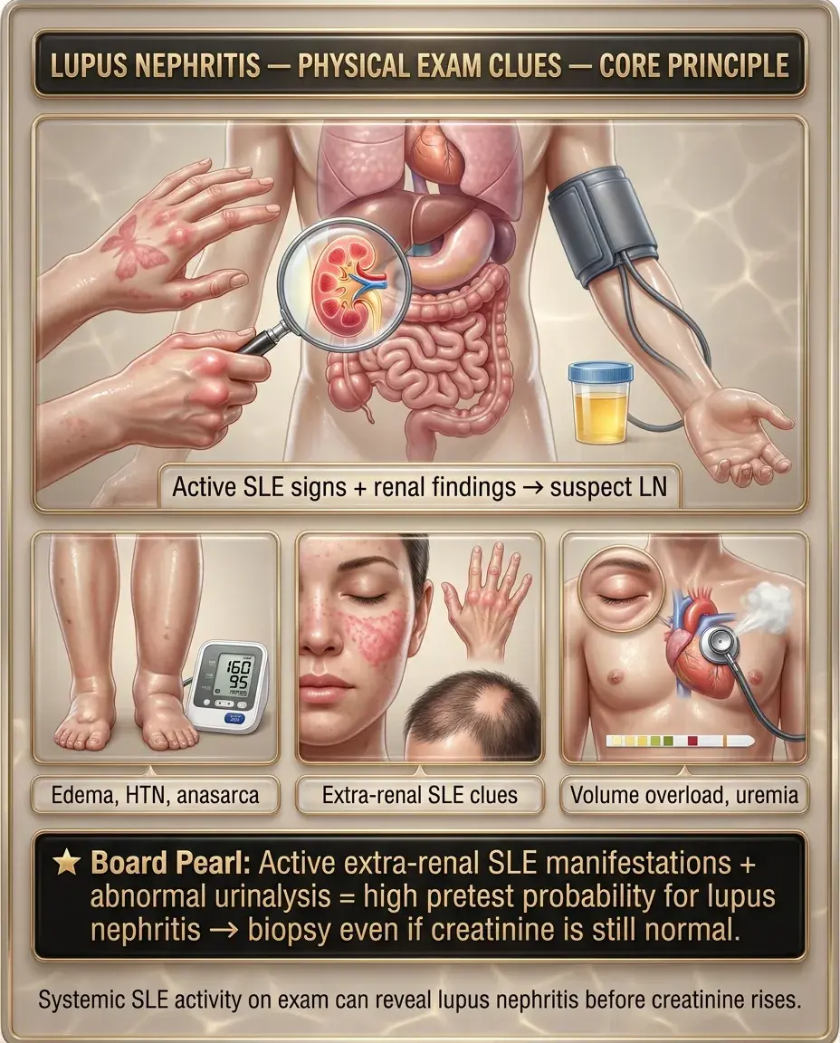
Physical exam often reflects both renal involvement and extra-renal SLE activity:

— Peripheral pitting edema (nephrotic syndrome)

— HTN — often new onset

— Anasarca in severe nephrotic presentations

— Malar (butterfly) rash, discoid lesions

— Oral/nasal ulcers (often painless)

— Arthritis — symmetric, non-erosive

— Alopecia — diffuse, non-scarring

— Serositis — pleural or pericardial rub

— Livedo reticularis (consider antiphospholipid syndrome overlap)

— Volume overload with pulmonary crackles

— Uremic features if progression to ESRD (pericardial rub, asterixis, nausea)

Board pearl: Active extra-renal SLE manifestations + abnormal urinalysis = high pretest probability for LN → biopsy even if creatinine is still normal.

Renal signs:
Extra-renal SLE signs supporting active disease:
Signs of advanced/chronic LN:
Solid White Background
Diagnostic Workup — Laboratory Evaluation

Initial labs:

Serologic markers:

Key distinction: ↑ anti-dsDNA + ↓ C3/C4 = serologic flare → correlates with active nephritis; persistently normal complements make active LN less likely.

Urinalysis: dysmorphic RBCs, RBC casts (hallmark of glomerulonephritis), WBC casts, proteinuria
Spot urine protein-to-creatinine ratio (UPCR): >0.5 g/g is threshold for LN diagnosis per ACR/EULAR
24-hour urine protein: gold standard but UPCR is acceptable and more practical
Serum creatinine + eGFR
Serum albumin: ↓ in nephrotic syndrome
CBC: may show cytopenias (lymphopenia, thrombocytopenia, hemolytic anemia)
Anti-dsDNA antibodies: ↑ titers correlate with renal disease activity
Complement C3 and C4: ↓ during active LN (consumed by immune complex formation)
ANA: virtually always positive but non-specific
Anti-Smith: highly specific for SLE but does not track disease activity
Solid White Background
Diagnostic Workup — Renal Biopsy and ISN/RPS Classification

Renal biopsy → mandatory for confirmed or suspected LN to guide therapy.

ISN/RPS Classification (2003, updated 2018):

Biopsy also reports activity index (reversible inflammation) vs chronicity index (irreversible scarring).

Board pearl: Class IV is the highest-yield class — most common, most aggressive, and requires the most intensive induction therapy. Always biopsy before starting treatment.

Class I — Minimal mesangial: normal LM, mesangial immune deposits on IF/EM → no specific treatment
Class II — Mesangial proliferative: mesangial hypercellularity → usually conservative management
Class III — Focal proliferative: <50% glomeruli involved → immunosuppression required
Class IV — Diffuse proliferative: ≥50% glomeruli involved → most common and most severe → aggressive immunosuppression
Class V — Membranous: subepithelial deposits → nephrotic syndrome predominant → immunosuppression
Class VI — Advanced sclerosing: ≥90% globally sclerosed glomeruli → irreversible → prepare for RRT
Solid White Background
First-Line Management — Induction Therapy

Goal: rapidly suppress active inflammation to preserve nephron mass.

For Class III/IV (± V) LN — aggressive induction:

— Preferred in most patients, especially African American/Hispanic (better response, fewer side effects than cyclophosphamide)

— Reserved for severe proliferative disease, rapidly progressive GN, or MMF failure

For pure Class V (membranous) LN:

Next best step: All LN patients should also receive hydroxychloroquine — ↓ flare risk, ↓ renal progression, ↓ thrombotic events, ↓ mortality.

Option 1: Mycophenolate mofetil (MMF) 2–3 g/day × 6 months
Option 2: IV cyclophosphamide (CYC) — low-dose Euro-Lupus protocol (500 mg q2 weeks × 6 doses) or NIH high-dose protocol
PLUS: Corticosteroids — IV methylprednisolone pulse (500–1000 mg × 3 days) → oral prednisone taper (start 0.5–1 mg/kg, taper over 3–6 months to ≤7.5 mg/day)
MMF + corticosteroids (first-line)
Calcineurin inhibitors (tacrolimus, cyclosporine) as alternatives
Solid White Background
Maintenance Therapy and Emerging Agents

After 6 months of induction → transition to maintenance (minimum 3–5 years, often longer):

Emerging / adjunctive agents:

Board pearl: Belimumab + standard of care is now a guideline-endorsed strategy for active LN — know this as a tested new addition to the treatment algorithm.

First-line maintenance: MMF 1–2 g/day (preferred) OR azathioprine 2 mg/kg/day
Continue low-dose prednisone (≤7.5 mg/day), with goal of complete steroid withdrawal
Continue hydroxychloroquine indefinitely
Belimumab (anti-BAFF/BLyS monoclonal antibody): FDA-approved add-on to standard therapy for active LN → ↓ renal flares, ↓ proteinuria; added during induction and continued through maintenance
Voclosporin (novel calcineurin inhibitor): FDA-approved add-on to MMF + steroids → ↑ complete renal response rates
Rituximab (anti-CD20): used off-label for refractory LN
Solid White Background
Supportive and Adjunctive Measures

Critical non-immunosuppressive therapies:

— ↓ intraglomerular pressure, ↓ proteinuria, renoprotective

— Target BP <130/80 mmHg

Next best step: Start ACEi/ARB + HCQ in every LN patient regardless of ISN/RPS class.

ACE inhibitor or ARB: mandatory if proteinuria >0.5 g/day
Hydroxychloroquine: continued in all SLE patients — renoprotective, ↓ flares, ↓ damage accrual
Statin therapy: for dyslipidemia (common in nephrotic syndrome)
Vitamin D supplementation: steroid-induced bone loss + SLE patients often deficient
Calcium supplementation + bone density monitoring on chronic steroids
Pneumococcal, influenza, COVID vaccination — immunocompromised state
Anticoagulation: consider if nephrotic syndrome with albumin <2.5 g/dL (↑ VTE risk) or concurrent antiphospholipid syndrome
SGLT2 inhibitors: emerging data for ↓ proteinuria and renoprotection in LN — increasingly recommended
Solid White Background
Special Populations — Pregnancy and Lupus Nephritis

LN and pregnancy:

Safe in pregnancy: HCQ (continue — ↓ flare risk), azathioprine, low-dose prednisone, tacrolimus

Contraindicated: MMF (teratogenic — neural tube defects, cleft lip), cyclophosphamide (teratogenic), ACEi/ARBs (fetotoxic — renal agenesis, oligohydramnios), methotrexate

— Switch MMF → azathioprine ≥3 months before conception

— Switch ACEi/ARB → nifedipine or labetalol for BP control

— LN flare: active sediment, ↑ anti-dsDNA, ↓ complement, extra-renal SLE activity

— Preeclampsia: ↑ LFTs, ↓ platelets, ↑ uric acid, normal/stable complements

Key distinction: ↓ C3/C4 + active sediment + ↑ anti-dsDNA → LN flare; ↑ uric acid + ↑ LFTs + ↓ platelets → preeclampsia.

Pre-conception counseling essential — plan pregnancy during remission (≥6 months quiescent disease)
Active LN ↑ risk of: preeclampsia, preterm birth, fetal loss, maternal renal deterioration
Medication adjustments:
Monitor: UPCR monthly, Cr, anti-dsDNA, C3/C4, CBC
Distinguish LN flare from preeclampsia:
Solid White Background
Special Populations — Pediatric, Elderly, and CKD

Pediatric LN:

Elderly patients:

Patients with pre-existing CKD:

Board pearl: In pediatric SLE, biopsy early — disease is more aggressive, and early treatment preserves long-term renal function.

More aggressive course than adult LN — higher proportion of Class IV
More likely to present with nephrotic syndrome + hypertension
Treatment: same agents (MMF, CYC, steroids) but cyclophosphamide gonadotoxicity is a major concern → MMF generally preferred
Growth monitoring: chronic steroids impair linear growth → steroid-sparing strategies essential
Late-onset SLE may have less renal involvement but higher treatment toxicity
↓ Immunosuppressive doses cautiously — ↑ infection risk
Monitor GFR closely — age-related decline + LN = accelerated CKD
Biopsy still indicated if new active sediment or ↑ proteinuria — need to distinguish active inflammation (treatable) from chronic scarring
High chronicity index on biopsy → ↓ likelihood of immunosuppressive benefit
Early nephrology co-management for CKD staging, AVF planning if approaching ESRD
Solid White Background
Complications and When to Escalate Care

— Creatinine doubling over days to weeks, active sediment, crescents on biopsy

Escalate: IV pulse methylprednisolone + IV cyclophosphamide ± plasmapheresis

— Consider ICU admission if concurrent pulmonary hemorrhage (lupus pneumonitis/DAH)

— Thromboembolism (renal vein thrombosis, PE) — albumin <2.5 g/dL → prophylactic anticoagulation

— Severe hyperlipidemia → statin therapy

— Infection risk compounded by immunosuppression + urinary Ig losses

— Failure to respond after 6 months of induction → repeat biopsy to reassess class and activity/chronicity

— Switch therapy: MMF failure → CYC (or vice versa)

— Add rituximab or voclosporin

— ~10–20% of LN patients despite therapy

— Prepare for dialysis or transplantation — LN patients do well post-transplant

— Disease quiescence usually required for ≥6 months before transplant listing

Next best step: Refractory LN after 6 months → repeat renal biopsy before changing therapy.

Rapidly progressive glomerulonephritis (RPGN):
Nephrotic syndrome complications:
Treatment-resistant LN:
Progression to ESRD:
Solid White Background
Thrombotic Microangiopathy and Overlap Syndromes

— ~30–40% of SLE patients have antiphospholipid antibodies

— APS nephropathy: thrombotic microangiopathy (TMA) on biopsy → arteriolar thrombosis, fibrin thrombi, cortical ischemia

— Distinct from immune complex LN — may coexist

— Treatment: anticoagulation (warfarin, target INR 2–3); immunosuppression alone insufficient

— SLE patients at ↑ risk for TTP/HUS

— Microangiopathic hemolytic anemia (schistocytes), thrombocytopenia, renal failure, fever, neurologic changes

Urgent: ADAMTS13 activity, peripheral smear → plasma exchange if TTP suspected

Key distinction: LN = immune complex GN (active sediment, low complement) vs APS nephropathy = thrombotic process (TMA on biopsy, positive aPL) vs TTP = MAHA + thrombocytopenia + ADAMTS13 <10%.

Antiphospholipid syndrome (APS) overlap:
Thrombotic thrombocytopenic purpura (TTP):
Solid White Background
Key Differentials — Glomerulonephritis in SLE Patients

Not all renal disease in SLE is lupus nephritis:

— NSAIDs → acute interstitial nephritis (AIN) or hemodynamic AKI

— Calcineurin inhibitors (tacrolimus) → ↑ Cr from vasoconstriction

— Look for eosinophiluria (AIN), recent med changes

Board pearl: Always biopsy to confirm LN rather than empirically treating — the differential is broad, and management differs dramatically based on histologic class.

Drug-induced nephrotoxicity:
IgA nephropathy: can coexist; mesangial IgA on biopsy
Diabetic nephropathy: if SLE + DM → nodular glomerulosclerosis on biopsy
Hypertensive nephrosclerosis: chronic HTN → arteriolosclerosis
Infection-associated GN: endocarditis, hepatitis B/C — check blood cultures, viral serologies
Solid White Background
Distinguishing LN from Other Causes of Nephrotic Syndrome

— LN Class V: "full house" immunofluorescence (IgG, IgA, IgM, C3, C1q) + subepithelial deposits

— Primary MN: IgG4-dominant, anti-PLA2R antibodies positive in ~70–80%, no C1q

Key distinction: C1q positivity + "full house" IF = lupus membranous

— FSGS: podocyte injury, segmental sclerosis, no immune deposits

— LN can have secondary FSGS-like changes in chronic disease

— Foot process effacement on EM without immune deposits

— Check ANCA if biopsy shows crescents without immune deposits

Next best step: "Full house" immunofluorescence on biopsy is virtually pathognomonic for lupus nephritis.

LN Class V (membranous) vs primary membranous nephropathy (MN):
LN vs focal segmental glomerulosclerosis (FSGS):
Minimal change disease (MCD): rare in SLE but can be NSAID-induced
ANCA-associated vasculitis: pauci-immune GN (no/scant immune deposits) — rare SLE overlap
Solid White Background
Monitoring and Follow-Up

Active disease monitoring (during induction):

Remission monitoring (during maintenance):

Response definitions:

Board pearl: Serologic flare (↑ anti-dsDNA, ↓ C3/C4) may precede clinical flare by weeks — use as an early warning to intensify monitoring.

UPCR or 24-hour urine protein: q1–3 months — target >50% ↓ by 6 months, complete remission (UPCR <0.5–0.7) by 12 months
Serum creatinine + eGFR: q1–3 months
Anti-dsDNA titers and C3/C4: q3 months — rising anti-dsDNA + falling complement predict flare
Urinalysis: monitor for RBC casts, active sediment
CBC: monitor for cytopenias (disease activity or drug toxicity)
LFTs: if on azathioprine or MMF
HCQ: annual ophthalmologic exam for retinal toxicity (after 5 years or earlier if risk factors)
UPCR + Cr + serologies: q3–6 months
Watch for flare: ↑ proteinuria, ↑ anti-dsDNA, ↓ complements, active sediment
Complete response: UPCR <0.5, normal Cr, inactive sediment
Partial response: ≥50% ↓ in proteinuria, stable/improved Cr
Solid White Background
Prevention of Flares and Long-Term Renal Outcomes

— ↓ renal flare rate by ~50%

— ↓ damage accrual, ↓ thrombosis, ↓ mortality

— Never discontinue unless retinal toxicity develops

— Chronic steroids → infection, osteoporosis, AVN, metabolic syndrome

Long-term outcomes:

Board pearl: Persistent proteinuria at 12 months (UPCR >0.7–1.0) predicts long-term renal decline — intensify therapy if remission targets are not met.

Hydroxychloroquine adherence: single most important preventive measure
Steroid minimization: target prednisone ≤5 mg/day or complete withdrawal
Blood pressure control: target <130/80; ACEi/ARB first-line
Avoid nephrotoxins: NSAIDs, aminoglycosides, IV contrast (with precautions)
Smoking cessation: smoking worsens renal outcomes in LN
Sunscreen and UV avoidance: UV triggers SLE flares → indirect renal protection
10-year renal survival ~85–90% with modern therapy
Poor prognostic factors: Class IV, high chronicity index, delayed treatment, African American race, non-adherence, persistent proteinuria >1 g at 12 months
Solid White Background
Ethical, Legal, and Patient Safety Considerations

— Cyclophosphamide is gonadotoxic → discuss oocyte/sperm cryopreservation before treatment, especially in young patients

— GnRH agonists (leuprolide) may be co-administered with CYC to ↓ ovarian failure risk

— MMF is teratogenic — ensure reliable contraception + pregnancy testing before initiation

— Risks: bleeding (most common), perinephric hematoma, AV fistula, infection

— Benefit: guides therapy and avoids empiric immunosuppression

— Biopsy is standard of care — declining biopsy limits ability to treat optimally

— Non-adherence is the leading modifiable risk factor for LN flare and ESRD

— Address barriers: cost (MMF vs generic), complexity, side effects, health literacy

— Involve social work, pharmacy, patient navigators in high-risk populations

— African American, Hispanic, and Asian patients have worse LN outcomes — partly biologic, partly due to social determinants of health

— Equitable access to nephrology, rheumatology, and newer agents (belimumab, voclosporin) must be addressed

Board pearl: Always counsel on fertility preservation before cyclophosphamide — this is a testable patient safety issue.

Fertility preservation:
Informed consent for renal biopsy:
Medication adherence:
Health disparities:
Solid White Background
High-Yield Associations and Rapid-Fire Clinical Facts

Board pearl: Wire-loop lesions = subendothelial immune deposits so thick they resemble capillary wall thickening on LM → Class IV LN.

Anti-dsDNA ↑ + C3/C4 ↓ → active lupus nephritis (most tested serologic pattern)
"Full house" IF (IgG, IgA, IgM, C3, C1q) → lupus nephritis
Class IV = diffuse proliferative = most common + most severe → wire-loop lesions on LM
RBC casts → glomerulonephritis (not UTI, not ATN)
Nephrotic syndrome + SLE → think Class V membranous LN
HCQ → ↓ flares, ↓ renal events, ↓ thrombosis, ↓ mortality → never stop
MMF preferred over CYC in African American patients with LN
Belimumab + voclosporin = newer FDA-approved add-ons for active LN
APS + LN → requires anticoagulation in addition to immunosuppression
Low serum albumin (<2.5 g/dL) in nephrotic LN → ↑ VTE risk → consider anticoagulation
ESRD from LN → transplant is preferred modality; disease recurrence in allograft is ~2–10%
Hypocomplementemia (↓ C3/C4) distinguishes LN from ANCA vasculitis (complement normal) and anti-GBM disease (complement normal)
Solid White Background
Board Question Stem Patterns
28F, SLE, malar rash, new edema, UA shows RBC casts, UPCR 3.2, ↑ anti-dsDNA, ↓ C3/C4 → Lupus nephritis → renal biopsy (next best step)
LN biopsy: ≥50% glomeruli with endocapillary proliferation, wire-loop lesions, "full house" IF → Class IV diffuse proliferative → IV methylprednisolone + MMF (or CYC)
African American woman, Class IV LN, choosing induction → MMF preferred over cyclophosphamide
SLE patient on MMF, wants to become pregnant → Switch MMF → azathioprine ≥3 months before conception; continue HCQ; stop ACEi
LN patient, 6 months induction, UPCR decreased from 4.0 → 1.5, Cr stable → Partial response → transition to maintenance MMF or azathioprine
SLE, nephrotic syndrome, albumin 1.8 g/dL, acute dyspnea, leg swelling → Renal vein thrombosis / PE → CT angiography + anticoagulation
SLE patient, ↑ proteinuria + ↑ anti-dsDNA + ↓ C3, currently on maintenance azathioprine → LN flare → repeat renal biopsy to guide re-induction
Pregnant SLE patient, 30 weeks, new proteinuria, ↓ C3/C4, active sediment → LN flare (not preeclampsia) → steroids + azathioprine
SLE, Class IV LN, refractory to MMF and CYC → Add rituximab or belimumab; re-biopsy to reassess
LN patient, stable remission, asks about stopping HCQ → Do not stop — HCQ ↓ flare risk and mortality; continue indefinitely
Solid White Background
One-Line Recap
Lupus nephritis is immune complex–mediated glomerulonephritis classified by renal biopsy into ISN/RPS Classes I–VI (Class IV diffuse proliferative is most common and severe), suspected when an SLE patient develops proteinuria >0.5 g/day, active urine sediment (RBC casts), or rising creatinine with serologic flare (↑ anti-dsDNA, ↓ C3/C4) and confirmed by mandatory renal biopsy showing "full house" immunofluorescence, managed with induction immunosuppression (MMF or cyclophosphamide + pulse then tapered corticosteroids), maintenance therapy (MMF or azathioprine for ≥3–5 years), universal hydroxychloroquine (never discontinue), ACEi/ARB for proteinuria and BP control, newer adjunctive agents (belimumab, voclosporin), fertility preservation counseling before cyclophosphamide, and serial monitoring of UPCR, creatinine, anti-dsDNA, and complement levels to detect flares early and guide treatment escalation.
Solid White Background
Previous Item
Next Item
bottom of page