top of page

Renal & Urinary

Chronic kidney disease: staging and management

Clinical Overview and When to Suspect Chronic Kidney Disease

Chronic kidney disease (CKD) → sustained structural or functional kidney abnormality for ≥3 months, defined by GFR <60 mL/min/1.73 m² or markers of kidney damage (albuminuria, abnormal sediment, structural abnormalities) regardless of GFR.

— Persistently ↑ serum creatinine or ↓ eGFR on two measurements ≥3 months apart

— Persistent albuminuria (UACR ≥30 mg/g on two of three specimens)

— Abnormal renal imaging: small echogenic kidneys, polycystic kidneys, cortical thinning

— Unexplained anemia, hyperkalemia, metabolic acidosis, hyperphosphatemia

Board pearl: A single ↑ creatinine does NOT equal CKD — chronicity (≥3 months) must be established. Always distinguish CKD from AKI by reviewing prior labs, kidney size on ultrasound (small = chronic), and presence of anemia/bone disease.

Classic patient: middle-aged or elderly adult with DM, HTN, or both — often discovered incidentally on routine labs
Suspect CKD when:
Solid White Background
Presentation Patterns and Key History

Key history to elicit:

— Duration of DM and glycemic control (diabetic nephropathy = #1 cause of CKD in U.S.)

— HTN duration and control (#2 cause — hypertensive nephrosclerosis)

— Family history: ADPKD, Alport syndrome, FSGS

— NSAID/nephrotoxin exposure (chronic interstitial nephritis)

— Recurrent UTIs, nephrolithiasis, BPH → obstructive uropathy

— Autoimmune diseases: SLE, vasculitis

— Prior AKI episodes — each episode ↑ risk of subsequent CKD

Next best step: In any patient with newly discovered ↓ eGFR, obtain prior creatinine values and renal ultrasound to assess chronicity and etiology.

Early CKD (stages 1–3a): typically asymptomatic — detected via screening labs
Moderate CKD (stages 3b–4): fatigue, nocturia (loss of concentrating ability), mild edema, ↑ BP
Advanced CKD/ESKD (stage 5): uremic symptoms — anorexia, nausea, dysgeusia (metallic taste), pruritus, restless legs, encephalopathy, pericarditis, peripheral neuropathy
Solid White Background
Physical Exam Findings and Signs of CKD Complications

Renal ultrasound clues:

— Small (<9 cm), echogenic kidneys → chronic parenchymal disease

— Normal or enlarged kidneys in CKD → consider DM nephropathy, amyloidosis, HIV-associated nephropathy, ADPKD

Board pearl: Normal-sized or large kidneys in a CKD patient should prompt consideration of diabetic nephropathy, infiltrative disease, or polycystic kidney disease — not all CKD kidneys are small.

Hypertension — both cause and consequence of CKD
Peripheral edema — nephrotic-range proteinuria or volume overload
Pallor — anemia of CKD (↓ erythropoietin)
Excoriations — uremic pruritus
Asterixis — uremic encephalopathy (late finding)
Pericardial friction rub — uremic pericarditis (indication for urgent dialysis)
Kussmaul respirations — metabolic acidosis compensation
Bilateral flank masses — autosomal dominant polycystic kidney disease (ADPKD)
Fundoscopic exam: diabetic/hypertensive retinopathy → correlates with nephropathy
AV fistula or peritoneal catheter in known ESKD patients
Solid White Background
Diagnostic Workup — Estimating GFR and Assessing Albuminuria

— Confirm with repeat eGFR ≥3 months later to establish chronicity

— Cystatin C–based eGFR useful when creatinine unreliable (extremes of muscle mass, amputation, cirrhosis)

— Spot urine albumin-to-creatinine ratio (UACR) — preferred screening test

— A1 (normal): <30 mg/g

— A2 (moderately increased, formerly "microalbuminuria"): 30–300 mg/g

— A3 (severely increased): >300 mg/g

— Confirm on ≥2 of 3 specimens over 3–6 months (transient causes: fever, exercise, UTI)

Key distinction: 24-hour urine protein collection is less practical but still used when spot UACR is discordant or when quantifying non-albumin proteinuria (e.g., light chains in myeloma).

eGFR: use CKD-EPI creatinine equation (preferred over MDRD and Cockcroft-Gault)
Albuminuria assessment:
CKD staging uses BOTH eGFR (G1–G5) AND albuminuria (A1–A3) → "heat map" determines prognosis and referral timing
Solid White Background
CKD Staging and Additional Workup

GFR stages:

Additional labs for all CKD patients:

Next best step: At CKD stage 3 and beyond, routinely monitor Ca²⁺, PO₄³⁻, PTH, Hgb, K⁺, and HCO₃⁻ — complications of CKD begin accumulating at this stage.

G1: eGFR ≥90 (kidney damage present but normal GFR)
G2: eGFR 60–89
G3a: eGFR 45–59
G3b: eGFR 30–44
G4: eGFR 15–29
G5: eGFR <15 (or dialysis)
BMP: Cr, BUN, K⁺, HCO₃⁻, Ca²⁺, phosphorus
CBC: anemia evaluation (begin checking at stage 3)
Intact PTH: secondary hyperparathyroidism screening (start at stage 3)
25-OH vitamin D: deficiency common
Lipid panel, HbA1c (if DM), hepatitis B/C serologies
Urinalysis with microscopy: dysmorphic RBCs → glomerular disease; WBC casts → interstitial nephritis
Renal ultrasound: size, symmetry, obstruction, cysts
Solid White Background
First-Line Management — Blood Pressure and Proteinuria Reduction

— Reduces intraglomerular pressure → slows progression

— Expect up to 30% ↑ in Cr after initiation — acceptable; do NOT stop unless >30% rise or hyperkalemia

— Monitor K⁺ and Cr within 1–2 weeks of starting/dose change

Do NOT combine ACEi + ARB (↑ hyperkalemia, AKI risk, no added benefit — ONTARGET trial)

— Now first-line add-on for CKD with albuminuria regardless of DM status (DAPA-CKD, EMPA-KIDNEY trials)

— Reduce progression to ESKD, cardiovascular events, and HF hospitalizations

— Mechanism: afferent arteriole vasoconstriction → ↓ intraglomerular pressure

— Expect initial ↓ eGFR ("dip") — acceptable; reflects hemodynamic effect

— Can initiate if eGFR ≥20 mL/min

Board pearl: The combination of ACEi/ARB + SGLT2i is the cornerstone of renoprotective therapy in proteinuric CKD.

BP target in CKD: <130/80 mmHg (per KDIGO 2021, especially with albuminuria)
ACEi or ARB — first-line for CKD with albuminuria (UACR ≥30 mg/g)
SGLT2 inhibitors (empagliflozin, dapagliflozin, canagliflozin):
Solid White Background
Additional Pharmacotherapy and Lifestyle Measures

— Indicated for T2DM-associated CKD with persistent albuminuria despite max ACEi/ARB + SGLT2i

— Reduces CKD progression and cardiovascular events (FIDELIO-DKN, FIGARO-DKD)

— Monitor K⁺ closely — risk of hyperkalemia

— HbA1c target ~7% (individualize based on hypoglycemia risk)

— Metformin: reduce dose at eGFR 30–45; discontinue at <30

— SGLT2i: dual benefit (glycemic + renoprotective)

— GLP-1 receptor agonists: cardiovascular and possible renal benefit

— Avoid glyburide in CKD (active metabolites → hypoglycemia)

— Sodium restriction: <2 g/day (reduces proteinuria and BP)

— Protein intake: 0.8 g/kg/day in non-dialysis CKD (avoid excessive restriction)

— Smoking cessation: accelerates CKD progression

— Regular exercise, weight management

Next best step: After starting ACEi/ARB + SGLT2i, add finerenone if diabetic CKD with persistent albuminuria.

Finerenone (nonsteroidal MRA):
Glycemic control in diabetic CKD:
Lifestyle:
Solid White Background
CKD-Mineral Bone Disorder (CKD-MBD) Management

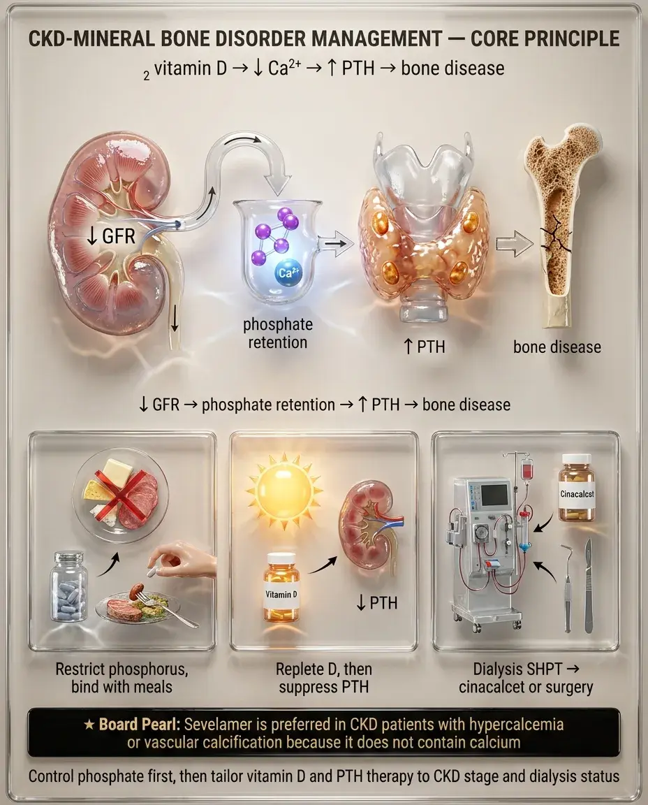
— Dietary phosphorus restriction (processed foods, dairy, cola)

— Phosphate binders with meals: calcium acetate, sevelamer (non-Ca²⁺ based — preferred if Ca²⁺ already ↑), lanthanum

— Target: normal serum phosphorus

— Replete 25-OH vitamin D deficiency with cholecalciferol/ergocalciferol

— Active vitamin D (calcitriol) or analogs (paricalcitol) for ↑ PTH in stages 4–5 when 25-OH D is replete

— In CKD G3–G5 (non-dialysis): trend PTH, treat if progressively rising

— On dialysis: target PTH roughly 2–9× upper limit of normal

— Cinacalcet (calcimimetic) for persistent secondary hyperparathyroidism on dialysis

— Parathyroidectomy for refractory tertiary hyperparathyroidism (autonomous PTH secretion with hypercalcemia)

Board pearl: Sevelamer is preferred in CKD patients with hypercalcemia or vascular calcification because it does not contain calcium.

Pathophysiology: ↓ GFR → phosphorus retention → ↓ 1,25-(OH)₂ vitamin D → ↓ Ca²⁺ → ↑ PTH (secondary hyperparathyroidism) → renal osteodystrophy
Phosphorus control:
Vitamin D:
PTH management:
Solid White Background
Special Populations — Diabetic CKD and Elderly

Diabetic nephropathy:

Elderly patients:

Board pearl: Absence of diabetic retinopathy in a T2DM patient with nephrotic-range proteinuria should raise suspicion for a non-diabetic glomerular disease → renal biopsy indicated.

Most common cause of CKD/ESKD in the U.S.
Natural history: hyperfiltration → microalbuminuria → overt proteinuria → declining GFR → ESKD
Screen annually with UACR starting 5 years after T1DM diagnosis, at diagnosis of T2DM
Biopsy not needed if classic presentation (progressive albuminuria, retinopathy present, >10 years DM)
Biopsy if: abrupt onset nephrotic syndrome, active urinary sediment (hematuria, RBC casts), rapid GFR decline, no retinopathy → suspect non-diabetic glomerular disease
Age-related GFR decline is common — eGFR 45–59 in an 80-year-old with no albuminuria may represent normal aging rather than progressive CKD
Drug dosing: renally cleared medications (DOACs, gabapentin, opioids) require adjustment
Conservative management (no dialysis) may be appropriate in frail elderly with multiple comorbidities — focus on symptom management and quality of life
Solid White Background
Special Populations — Pregnancy and Pediatric CKD

Pregnancy in CKD:

Pediatric CKD:

Board pearl: ACEi/ARB must be stopped immediately in any CKD patient who becomes pregnant — teratogenic effects occur in all trimesters.

CKD ↑ risk of pre-eclampsia, preterm birth, fetal growth restriction, accelerated GFR decline
ACEi/ARB — absolutely contraindicated (teratogenic: renal agenesis, oligohydramnios)
Switch to labetalol, nifedipine, or methyldopa for BP control
SGLT2i, finerenone — discontinue preconception (insufficient safety data)
Low-dose aspirin (from 12 weeks) for pre-eclampsia prevention
Close monitoring: monthly eGFR, UACR, BP; fetal surveillance
Referral to high-risk obstetrics and nephrology co-management
Common causes: congenital anomalies of the kidney and urinary tract (CAKUT), focal segmental glomerulosclerosis, reflux nephropathy
Growth failure is a major concern — manage with recombinant growth hormone if refractory to nutritional optimization
Early referral to pediatric nephrology
Vaccination schedule: complete immunizations before any transplant (live vaccines contraindicated post-transplant)
Solid White Background
Complications — Anemia, Acidosis, and Hyperkalemia

Anemia of CKD:

— IV iron preferred in dialysis patients; oral or IV in non-dialysis CKD

— Start only after iron repletion if Hgb <10 g/dL

— Target Hgb 10–11.5 g/dL — do NOT target >13 (↑ cardiovascular events, stroke — CHOIR/TREAT trials)

— HIF-prolyl hydroxylase inhibitors (roxadustat): oral alternative

Metabolic acidosis:

Hyperkalemia:

Board pearl: Always correct iron deficiency before starting ESAs — iron-deficient erythropoiesis renders ESAs ineffective.

Normocytic, normochromic; primarily from ↓ erythropoietin production
Evaluate at eGFR <60: check Hgb, iron studies (ferritin, TSAT)
Treat iron deficiency FIRST: target ferritin >100 ng/mL, TSAT >20% (non-dialysis); ferritin >200, TSAT >20% (dialysis)
Erythropoiesis-stimulating agents (ESAs): epoetin alfa, darbepoetin alfa
↓ NH₄⁺ excretion → non-anion-gap metabolic acidosis early; ↑ anion gap in advanced CKD
Supplement with oral sodium bicarbonate to maintain HCO₃⁻ ≥22 mEq/L — slows CKD progression
↓ renal K⁺ excretion, worsened by ACEi/ARB, MRAs, high-K⁺ diet
Chronic management: dietary restriction, loop diuretics, K⁺ binders (patiromer, sodium zirconium cyclosilicate) — these enable continued use of RAASi
Solid White Background
When to Refer and Urgent Dialysis Indications

Nephrology referral:

Dialysis preparation:

Emergent dialysis indications (mnemonic-free approach):

Board pearl: Uremic pericarditis is an absolute indication for urgent dialysis regardless of other parameters.

eGFR <30 (stage 4) — plan for renal replacement therapy (RRT)
Rapidly declining eGFR (>5 mL/min/year)
Persistent albuminuria ≥300 mg/g despite optimal ACEi/ARB
Suspected glomerulonephritis (active sediment, hematuria + proteinuria)
Refractory HTN, hyperkalemia, or acidosis
CKD of unknown etiology
Discuss modalities at eGFR <20: hemodialysis, peritoneal dialysis, preemptive transplant
AV fistula creation 6 months before anticipated need (time for maturation)
Hepatitis B vaccination — check titer; CKD patients have impaired response → may need higher dose
Refractory hyperkalemia (K⁺ >6.5 with ECG changes, unresponsive to medical therapy)
Severe metabolic acidosis (pH <7.1, refractory to bicarbonate)
Volume overload refractory to diuretics (pulmonary edema)
Uremic pericarditis or encephalopathy
Certain toxic ingestions (methanol, ethylene glycol, lithium, salicylates)
Solid White Background
Key Differentials — CKD vs AKI vs AKI on CKD

Distinguishing CKD from AKI:

AKI superimposed on CKD:

Other causes of ↑ Cr without true GFR decline:

Key distinction: A patient with CKD whose Cr rises acutely after NSAID or contrast exposure has AKI on CKD — the acute component may be reversible.

Prior labs showing chronically ↑ Cr → CKD
Small, echogenic kidneys on US → CKD
Anemia, hyperphosphatemia, ↑ PTH → suggest chronicity
Broad, waxy casts on UA → CKD
AKI: abrupt Cr rise (within hours–days), normal prior labs, normal-sized kidneys
CKD patients are highly susceptible to AKI from: volume depletion, contrast, NSAIDs, obstruction
Key clue: acute Cr rise above an established elevated baseline
Management: treat the reversible AKI component; may not return to prior baseline
Trimethoprim, cimetidine → block tubular Cr secretion → ↑ Cr with stable GFR
Increased muscle mass or high-protein meal → transiently ↑ Cr
Cystatin C–based eGFR helps clarify
Solid White Background
Differentiating Causes of CKD — Glomerular vs Tubulointerstitial vs Vascular

Glomerular:

Tubulointerstitial:

Vascular:

Board pearl: Sudden ↑ Cr after starting ACEi in bilateral renal artery stenosis (or unilateral in a solitary kidney) → bilateral renovascular disease. Hold ACEi/ARB and obtain imaging (duplex US, MRA, or CTA).

Proteinuria (often >3.5 g/day if nephrotic), active sediment (dysmorphic RBCs, RBC casts)
DM nephropathy, FSGS, membranous, IgA nephropathy, lupus nephritis
Biopsy for diagnosis when etiology unclear
Bland sediment, WBC casts, mild proteinuria (<1–2 g/day)
Causes: chronic NSAID/analgesic use, lithium, lead, reflux nephropathy, Sjögren
May present with tubular dysfunction: type 4 RTA, concentrating defect
Hypertensive nephrosclerosis: long-standing HTN, LVH, retinopathy, mild proteinuria
Renovascular disease (atherosclerotic RAS): asymmetric kidneys, flash pulmonary edema, ↑ Cr with ACEi
Cholesterol embolism: post-catheterization, livedo reticularis, blue toes, eosinophilia, ↓ complement
Solid White Background
Preventive Care and Screening in CKD

— Serum creatinine → eGFR

— Spot UACR

— Statin therapy: moderate-to-high intensity for CKD stages 1–4 (atorvastatin preferred — not renally cleared)

— Statin benefit less clear in dialysis patients (4D, AURORA trials neutral) — continue if already on statin but no need to initiate

— Aspirin: individualized based on ASCVD risk

— Influenza (annually), pneumococcal (PCV20 or PCV15 + PPSV23), hepatitis B (high-dose formulation)

— COVID-19 vaccination + additional doses (immunocompromised protocol)

— CKD patients have impaired vaccine responses → check hepatitis B titers, revaccinate if non-responder

Board pearl: Gadolinium-based contrast is contraindicated in eGFR <30 due to risk of nephrogenic systemic fibrosis — use group II agents if absolutely necessary.

Screen for CKD annually in high-risk populations: DM, HTN, cardiovascular disease, family history of CKD, AKI history, obesity
Cardiovascular risk reduction (CKD = coronary risk equivalent):
Vaccinations:
Avoid nephrotoxins: NSAIDs, aminoglycosides, iodinated contrast (use with hydration if necessary), gadolinium (risk of nephrogenic systemic fibrosis if eGFR <30)
Solid White Background
Monitoring and Follow-Up Schedule

— G1–G2, A1: annual eGFR, UACR, BP

— G3a, A1–A2: every 6–12 months

— G3b–G4: every 3–6 months

— G5/dialysis: monthly or more frequent

— eGFR, UACR, BMP (K⁺, HCO₃⁻, Cr)

— BP assessment and medication reconciliation

— Assess medication dosing for renal clearance

— CBC for anemia

— Ca²⁺, PO₄³⁻, intact PTH (frequency ↑ with advancing stage)

— 25-OH vitamin D annually

— Lipids, HbA1c (if DM)

Next best step: If eGFR declining faster than expected, reassess medication adherence, check for ongoing nephrotoxin exposure, evaluate for superimposed AKI, obstruction, or new glomerular disease.

Frequency based on CKD stage and albuminuria category:
At each visit:
Periodic monitoring (every 3–6 months for stages 3–5):
Track eGFR slope: decline >5 mL/min/year is rapid progression → intensify therapy, investigate reversible causes
Solid White Background
Ethical, Legal, and Patient Safety Considerations

— Conservative management (no dialysis) is a valid option, especially in frail elderly with poor functional status and multiple comorbidities

— Discuss prognosis honestly — address goals of care early (ideally at CKD stage 4)

— Palliative care referral for symptom management in those choosing conservative care

— Document wishes regarding dialysis withdrawal, resuscitation status

— Dialysis withdrawal is the second most common cause of death in ESKD patients — counsel families

— Many drugs require dose adjustment or avoidance in CKD (DOACs, metformin, gabapentin, opioids)

— Pharmacy reconciliation at every visit

— NSAIDs: avoid in CKD stages 3–5 (accelerate progression, cause AKI, hyperkalemia)

— CKD disproportionately affects Black, Hispanic, and Native American populations

— eGFR equations have removed race coefficients (CKD-EPI 2021) to reduce disparities in diagnosis and referral

— Ensure equitable access to transplant evaluation

Board pearl: The 2021 CKD-EPI equation no longer includes a race coefficient — this change was made to ensure equitable diagnosis and referral for all patients.

Shared decision-making for dialysis initiation:
Advance directives:
Medication safety:
Health equity:
Solid White Background
High-Yield Associations and Rapid-Fire Clinical Facts

Board pearl: Episodic gross hematuria during URIs with underlying CKD → think IgA nephropathy; post-infectious GN occurs 2–3 weeks AFTER infection.

DM nephropathy: #1 cause of ESKD in U.S.; Kimmelstiel-Wilson nodules on biopsy
HTN nephrosclerosis: #2 cause; arteriolar hyalinosis, interstitial fibrosis
ADPKD: large kidneys bilaterally; associated with cerebral aneurysms, hepatic cysts, MVP
IgA nephropathy: episodic gross hematuria 1–2 days after URI ("synpharyngitic")
Alport syndrome: hematuria + sensorineural hearing loss + ocular abnormalities; X-linked most common
ACEi/ARB: ↓ proteinuria and CKD progression — tolerate Cr ↑ ≤30%
SGLT2i: renoprotective in DM and non-DM CKD → initial eGFR "dip" is expected
ESA target Hgb: 10–11.5 g/dL (never >13)
Oral NaHCO₃ to keep HCO₃⁻ ≥22 → slows CKD progression
Patiromer / sodium zirconium cyclosilicate → K⁺ binders enabling continued RAASi use
AV fistula preferred over graft or catheter → lower infection/thrombosis rates
Calciphylaxis: painful necrotic skin lesions in dialysis patients with ↑ Ca²⁺ × PO₄³⁻ product
Solid White Background
Board Question Stem Patterns
55M, DM × 15 years, Cr 1.8, UACR 450 mg/g, retinopathy present → Diabetic nephropathy → ACEi/ARB + SGLT2i + optimize glycemic control
CKD patient, Cr rises from 1.5 → 1.9 after starting lisinopril (27% increase) → Expected hemodynamic effect — continue ACEi, recheck in 1–2 weeks
CKD G4, Hgb 8.5, ferritin 50, TSAT 15% → Iron deficiency → IV iron FIRST, then reassess need for ESA
70F, CKD G3b, K⁺ 5.8, on lisinopril → Start patiromer (K⁺ binder) to maintain ACEi therapy
CKD G5, asterixis, pericardial friction rub → Uremic pericarditis → urgent dialysis
T2DM, eGFR 35, UACR 200 mg/g, on max-dose losartan + dapagliflozin → Add finerenone for additional renoprotection
CKD patient, PO₄ 6.5, Ca²⁺ 10.8, PTH elevated → Start sevelamer (non-calcium phosphate binder)
80F, CKD G5, frail, multiple comorbidities, family asks about dialysis → Discuss conservative management; palliative care referral
Patient with CKD, bilateral small kidneys on US, anemia, ↑ PTH → Findings confirm chronicity — this is CKD, not AKI
Post-contrast Cr rise in CKD patient → AKI superimposed on CKD — supportive care, IV fluids, avoid further nephrotoxins
Solid White Background
One-Line Recap
CKD is defined by eGFR <60 mL/min/1.73 m² or kidney damage markers for ≥3 months, staged by both GFR (G1–G5) and albuminuria (A1–A3), most commonly caused by diabetes and hypertension, managed with ACEi/ARB as first-line for proteinuria reduction, SGLT2 inhibitors as disease-modifying add-on therapy regardless of DM status, finerenone for diabetic CKD with persistent albuminuria, BP target <130/80, oral NaHCO₃ to maintain HCO₃⁻ ≥22, iron repletion before ESAs with Hgb target 10–11.5 (never >13), phosphate binders and vitamin D for CKD-MBD, K⁺ binders to enable continued RAASi use, nephrology referral at eGFR <30 for RRT planning (AV fistula 6 months ahead), urgent dialysis for refractory hyperkalemia/acidosis/volume overload or uremic pericarditis/encephalopathy, and shared decision-making regarding dialysis versus conservative management in frail elderly patients.
Solid White Background
Previous Item
Next Item
bottom of page